PANCE Blueprint GI and Nutrition (9%)

Ischemic bowel disease

Patient will present as  a 71-year-old male with a history of atrial fibrillation with a sudden onset of severe abdominal pain occurring 10 minutes after eating. Physical exam findings are relatively benign, and the patient has only minimal pain with palpation of the abdomen. Stool guaiac is positive, and blood tests reveal leukocytosis with an elevated lactate, amylase, and LDH.

What is the most common vessel blocked with intestinal ischemia?
Superior mesenteric artery
Severe abdominal pain 30 minutes after a meal should make you think of what diagnosis?
Intestinal ischemia

Ischemic bowel disease occurs when an artery that supplies blood becomes blocked or narrowed. This can affect the small bowel or the large bowl. 

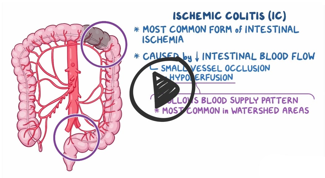
  • There are several possible causes of ischemic bowel disease, including blockage in the arteries due to a tumor or blood clot, narrowing of the arteries supplying blood to the bowel from atherosclerosis, hernia, volvulus, and intussusception. It can also occur due to a nonocclusive event like low blood pressure throughout the body (shock).

Types of intestinal ischemia

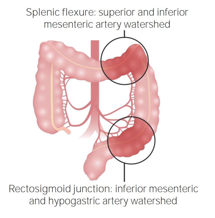
  • Ischemic colitis (ischemia of the colon): hypoperfusion of the large bowel
  • Acute mesenteric ischemia: acute loss of blood flow to the small intestine
  • Chronic mesenteric ischemia: constant or episodic hypoperfusion of the small intestine
Ischemic colitis (ischemia of the colon)

  • The most common type of intestinal ischemia (approximately 60%–70%)
  • Rapid onset of mild abdominal pain over the affected bowel
  • Mild rectal bleeding and bloody diarrhea, usually within 24 hours of abdominal pain
  • Caused by decreased intestinal blood flow - most common in the watershed areas of the colon (splenic flexure and rectosigmoid junction)
  • Ischemic colitis occurs mainly in older people (> 60) and is thought to be caused by small-vessel atherosclerosis. It can also be a complication of abdominal aortic aneurysm repair.
  • Risk factors for colonic ischemia include hypertension, diabetes Mellitus, atrial fibrillation, and aortic surgery, during which the aorta and its branches might be temporarily clamped, which can cause hypoperfusion of the colon
  • Labs reveal elevated white blood cells and lactate
  • CT scan with IV and oral contrast - signs of perforation include free air in the abdomen.
  • Bowel wall thickening and submucosal edema appear as “thumbprinting” on radiograph or CT imaging
Acute mesenteric ischemia (ischemia of the small intestine)

  • Sudden onset of acute and severe abdominal pain out of proportion to the exam
  • Board review questions will likely present with a patient with a-fib, MI, or CHF
  • X-ray shows “thumb-printing” of the small bowel or right colon due to submucosal bleeding
  • The superior mesenteric artery is the most common site
  • 1 in 1,000 of all hospital admissions
  • 1–2% of all admissions for abdominal pain: Most cases occur in patients >50 yr. Mortality as high as 60–70%, particularly if diagnosis/presentation delayed >24 hr.
  • It has also been described in young patients using oral contraceptive pills or illicit drugs
  • Aggressive pursuit of diagnosis is mandatory. Early surgical evaluation for emergent operative intervention is mandatory.

Chronic mesenteric ischemia (ischemia of the small intestine)

  • Postprandial epigastric pain usually occurs within the first hour after eating
  • Pain will resolve in 2-3 hours
  • The superior mesenteric artery is the most common site
  • Mesenteric angina
  • Usually will present as recurrent cramping with postprandial pain in a patient with a history of PVD, smoking, or DM

Diagnosis is made by CT or colonoscopy, angiography, or magnetic resonance angiography

  • Mesenteric angiography is considered the (gold standard)
  • Abdominal X-ray shows “thumb-printing” of small bowel or right colon due to submucosal bleeding

Treatment for both acute and chronic mesenteric ischemia is revascularization

osmosis Osmosis
Picmonic
Mesenteric ischemia

IM_MED_MesentericIschemia_V1.7_ASSETS

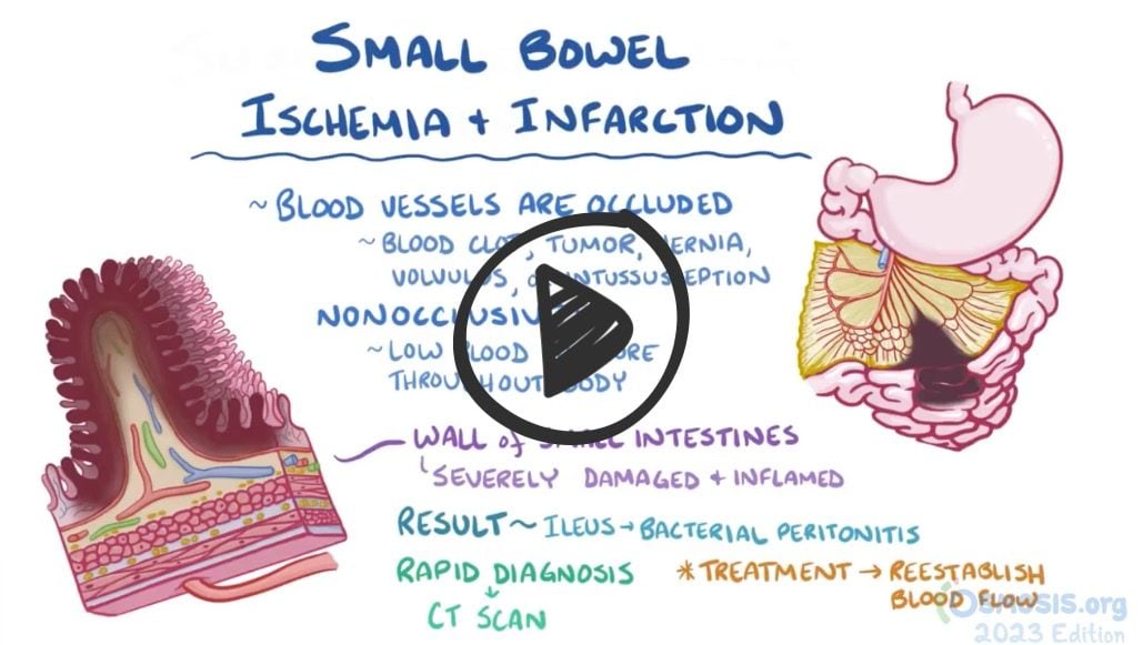
Mesenteric ischemia is a condition caused by insufficient blood flow to the small intestine. Poor blood flow is caused by thrombotic or embolic occlusion of the arteries that feed the small intestine, most commonly the proximal superior mesenteric artery (SMA). This lack of perfusion causes bowel ischemia, and subsequent necrosis if left untreated. Hallmark findings are severe abdominal pain that is out of proportion to their physical examination, nausea, vomiting, and bloody stools. Laboratory testing shows metabolic acidosis with elevated lactate, and abdominal x-ray and CT angiography may show bowel wall thickening or edema, signs of obstruction, or air within the bowel wall. Treatment includes fluids, broad spectrum antibiotics, anticoagulation, and emergent surgical exploration in severe cases.

Play Video + Quiz

Question 1
Thumb printing, a radiographic sign, is seen in all of the following conditions except
A
Crohn disease
Hint:
See C for explanation
B
Ulcerative colitis
Hint:
See C for explanation
C
Appendicitis
D
Ischemic colitis
Hint:
See C for explanation
Question 1 Explanation: 
Thumbprinting is a radiographic sign of large bowel thickening caused by edema, related to an infective or inflammatory process. It is not seen in appendicitis. It is seen in all other options.
Question 2
A 65-year old man who’s being managed for arrhythmia presented at the emergency with sudden onset severe abdominal pain 10 minutes after eating and bleeding per rectum. He had nausea and had vomited thrice prior to presentation. He also confessed to have been non-drug compliant. Physical examination findings were out of proportion with the abdominal pain. What is this patient’s most likely diagnosis?
A
Acute mesenteric ischemia
B
Diverticulitis
Hint:
Physical examination would reveal a palpable mass at the left lower quadrant of the abdomen.
C
Ulcerative colitis
Hint:
Symptoms onset is typically insidious. There are also extraintestinal manifestations. Abdominal pain doesn’t typically occur following feeding. Cardiac disease is not a risk factor.
D
Crohn disease
Hint:
Abdominal pain doesn’t typically occur following feeding. There are extraintestinal manifestations, and a mass may be felt at the right lower quadrant.
Question 2 Explanation: 
AMI typically presents as sudden onset abdominal pain occurring 10 -30 minutes after feeding. It is associated with bleeding per rectum with or without diarrhea. Physical examination findings is usually disproportionate with abdominal pain. It occurs in the elderly and those that are at risk of emboli formation.
Question 3
A 62-year old woman, a known hypertensive and diabetic patient, presents at the clinic with 4-months history of dull epigastric pain which usually occurs 30 minutes after feeding. This has lead to her having fear to eating. She has lost 11lb in the past 7 weeks. There is also nausea, vomiting and diarrhea. She also complained of what seemed like intermittent claudication. Physical examination reveals signs of malnutrition, diffuse mild abdominal tenderness with no rebound and guarding, abdominal bruit, shiny skin on both legs, hair loss on both legs and feet, and weak dorsalis pedis pulse bilaterally. What is her most likely diagnosis?
A
Acute mesenteric ischemia
Hint:
presents as sudden onset abdominal pain with rectal bleeding. Presentation is usually acute.
B
Peptic ulcer disease
Hint:
Presents as periodic epigastric pain. Pain may be made worse by certain types of food.
C
Chronic mesenteric ischemia
D
Gastric cancer
Hint:
Epigastric pain is not associated with feeding
Question 3 Explanation: 
Chronic mesenteric ischemia typically presents as stated above. Patient with atherosclerosis or at risk of having atherosclerosis are predisposed to CMI. Intermittent claudication, shiny skin on both legs, hair loss on both legs and feet, and weak dorsalis pedis pulse bilaterally are signs of peripheral vascular disease which is due to atherosclerosis. Hypertension and Diabetes are risk factors for atherosclerosis.
Question 4
Which of the following is not a pharmacological option for acute mesenteric ischemia (AMI)?
A
Papaverine
Hint:
Intra-arterial infusion of papaverine through the angiography catheter at the affected vessel is useful for all arterial forms of AMI.
B
Urokinase
Hint:
A thrombolytic, infused through the angiography catheter can be live saving for selected patient with acute mesenteric arterial embolism.
C
Heparin
Hint:
Heparin anticoagulation is the main therapy for mesenteric venous thrombosis.
D
Prednisolone
Question 4 Explanation: 
Prednisolone (a corticosteroid) plays no role in the pharmacological management of AMI.
Question 5
Surgical management for chronic mesenteric ischemia includes all of the following except
A
Angioplasty with stenting
Hint:
See D for explanation
B
Angioplasty without stenting
Hint:
See D for explanation
C
Revascularization
Hint:
See D for explanation
D
All of the above are appropriate surgical management options
Question 5 Explanation: 
All of the above are appropriate surgical management options
There are 5 questions to complete.
List
Return
Shaded items are complete.
12345
Return

References: Merck Manual · UpToDate

Irritable bowel syndrome (Prev Lesson)
(Next Lesson) Large Bowel Obstruction
Back to PANCE Blueprint GI and Nutrition (9%)

NCCPA™ CONTENT BLUEPRINT