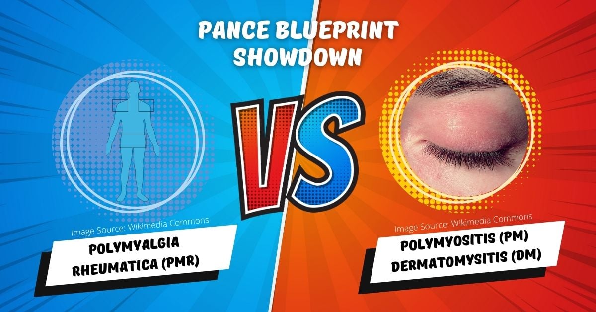
The table below provides a concise overview of these three conditions, helping PA students and practicing PAs quickly differentiate between them:
Blueprint Comparisons you need to know: (Polymyalgia Rheumatica (PMR) vs. Polymyositis (PM) vs. Dermatomyositis (DM)
| Aspect | Polymyalgia Rheumatica (PMR) | Polymyositis (PM) | Dermatomyositis (DM) |
| Essentials | Polymyalgia rheumatica (PMR) is an idiopathic inflammatory disorder that causes muscle pain and stiffness, especially in the shoulders and hips. It can be associated with giant cell arteritis. Proximal articular structures (bursae and tendons) that are mainly affected in PMR | Polymyositis is a chronic autoimmune inflammatory disease of muscle causing symmetrical, proximal muscle weakness | Dermatomyositis is an autoimmune myopathy characterized by symmetric proximal muscle weakness AND characteristic cutaneous findings |
| Age of Onset | Almost exclusively a disease of adults over the age of 50, with a prevalence that increases progressively with advancing age | Adults 30-50 years old, can occur at any age | Adults 40-60 years old, can occur in children |
| Primary Symptoms | Muscle stiffness, pain in shoulders and hips, morning stiffness lasting more than 45 minutes | Progressive muscle weakness, particularly in proximal muscles (e.g., shoulders, hips) | Muscle weakness, distinctive skin rash (heliotrope rash, Gottron's papules) |
| Associated Conditions | Often associated with Giant Cell Arteritis | It may be associated with other autoimmune conditions like Scleroderma, Lupus, etc. | Associated with malignancies in adults, calcinosis in children |
| Laboratory Findings | Elevated ESR and CRP, normal CK
Autoantibodies are usually negative in PMR |
Elevated CK, Aldolase, LDH, AST, and ALT
May have specific autoantibodies (anti-Jo-1) |
Elevated CK, positive ANA, specific autoantibodies (e.g., anti-Mi-2, anti-Jo-1) |
| Muscle Involvement | No actual muscle weakness; pain and stiffness are predominant | True muscle weakness, often symmetrical and progressive | Muscle weakness, often in proximal muscles, and skin involvement |
| Treatment | Corticosteroids (e.g., Prednisone) are mainstay of treatment | Corticosteroids, along with other immunosuppressants such as Methotrexate or Azathioprine | Corticosteroids, immunosuppressants, and treatment for skin involvement |
| Diagnosis | Clinical diagnosis supported by lab findings and response to steroids
A muscle biopsy for polymyalgia rheumatica (PMR) typically shows no evidence of disease |
Muscle biopsy is often required for a definitive diagnosis | Muscle biopsy, skin biopsy for definitive diagnosis |
| Prognosis | Generally good with treatment, often resolves within 1-3 years | Chronic and progressive response to treatment varies and may lead to significant disability | Variable: skin symptoms respond well to treatment, muscle weakness varies, and increased cancer risk in adults |
Understanding these differences is key for effective diagnosis and treatment and is essential for those preparing for the PANCE, PANRE, and EOR exams. As you study these conditions, remember to focus on their unique symptoms, lab findings, associated conditions, and treatment approaches to ensure a comprehensive understanding.
Polymyositis, Dermatomyositis, and Polymyalgia Rheumatica are covered under your PANCE/PANRE Musculoskeletal Blueprint as well as on the Internal Medicine EOR Topic List under the category of Rheumatologic Disorders.
For a more in-depth analysis of each of these conditions, along with concise clinical vignettes, flashcards, and quizzes, view the individual lesson at:
- Polymyositis (PM) and Dermatomyositis (DM)
- Polymyalgia Rheumatica (PMR)
- PANCE/PANRE Rheumatologic Conditions (PEARLS)
Stay tuned for our next PANCE Blueprint Showdown, where we continue to dissect essential Blueprint comparisons you need to know.
Happy studying!
Stephen Pasquini PA-C


