PANCE Blueprint EENT (7%)

PANCE Blueprint EENT (7%)
Featured

PANCE Blueprint EENT (7%)

Follow along with the NCCPA™ PANCE and PANRE EENT Content Blueprint

Lessons

  1. EENT 109 Question Comprehensive Exam (Members Only)

    Comprehensive PANCE/PANRE EENT Blueprint Exam
        • Viral: copious watery discharge, scant mucoid discharge. Adenovirus (most common). Self-limiting associated with URI.
        • Bacterial:  Pt will present with purulent (yellow) discharge, crusting, usually worse in the morning. May be unilateral.
          • S. pneumonia, S. aureus – acute mucopurulent
          • M. catarrhalis, Gonococcal – copious purulent discharge, in a patient who is not responding to conventional treatment
          • Chlamydia– newborn, giemsa stain - inclusion body, scant mucopurulent discharge
        • Allergic: red eyes, itching and tearing, usually bilateral, cobblestone mucosa on the inner/upper eyelid.
      1. Blurred vision over months or years, halos around lights. Clouding of the Lens (versus clouding of cornea = glaucoma). Fundoscopy "black on red background."
      2.  Contact lense wearers, caused by deep infection in the cornea by bacteria, viruses or fungi. White spot on surface of cornea that stains with fluorescein: round "ulceration" versus "dendritic" pattern like herpes
      3. Keratitis

        Keratitis is a condition in which the cornea becomes inflamed and is often marked by pain, impaired eyesight, photophobia, and red eye
        • Infectious keratitis can be caused by bacteria, viruses, fungi, or parasites
        • Viral infection of the cornea is often caused by the herpes simplex virus which causes a dendritic ulcer with fluorescein stain
        • Bacterial infectious keratitis - improper contact lens wear is the largest risk factor and commonly presents with a corneal opacity or infiltrate (typically a round white spot)
        • Treat herpes keratitis with topical antiviral
        • Bacterial keratitis requires urgent ophthalmological referral and prompt initiation of topical bactericidal antibiotics (ideally after obtaining cultures)
      4. Elevated, superficial, fleshy, triangular-shaped “growing” fibrovascular mass (most common in inner corner/nasal side of the eye). Only surgically remove when vision is affected
        • Dacryoadenitis is inflammation of the nasolacrimal duct or the nasolacrimal gland (supratemporal)
        • Dacryocystitis is an infectious obstruction of the nasolacrimal duct (inferomedial region)
      1. Eyelid changes: crusting, greasy, scaling, red-rimming of eyelid and eyelash flaking along with dry eyes and associated seborrhea and rosacea
      2. A chalazion is a sterile painless (non-infectious) granuloma of the internal meibomian sebaceous gland, painless "cold" lid nodule
      3. Ectropion (eversion of the eyelid) occurs when the eyelid turns outward exposing the palpebral conjunctiva, conjunctiva will appear red from air exposure and inflammation
      4. Entropion (inversion of an eyelid) occurs when the eyelid turns inward. It is most commonly caused by age-related tissue relaxation, surgical correction is definitive
      5. Painful, warm (hot), swollen red lump on the eyelid (different from a chalazion which is painless) Think “H” for Hot = Hordeolum. Most common organism S.  aureus.
        • Down/upbeat: CNS dysfunction
        • Vestibular (horizontal): labyrinth or vestibular nerve dysfunction
        • Gaze-evoked: most common and often benign
      1. Acute inflammation and demyelination of the optic nerve leading to acute monocular vision loss/blurriness and pain on extraocular movements. Typically occurs over hours or days. Fundoscopy: inflammation of the optic disc. Associated with multiple sclerosis.
      2. Optic disc swelling that is caused by increased intracranial pressure. The swelling is usually bilateral and can occur over a period of hours to weeks. Causes include brain tumor/abscess, meningitis, cerebral hemorrhage, encephalitis, pseudotumor cerebri. Immediate neuroimaging to rule out mass lesion, then CSF analysis. Treat underlying cause.
      1. Decreased extraocular movement, pain with movement of the eye and proptosis, signs of infection. Often associated with sinusitis. Occurs more often in children than adults. Hospitalization and IV broad-spectrum antibiotics.
      1. Gradual painless loss of central vision. The macula is responsible for central visual acuity which is why macular degeneration causes gradual central field loss. Metamorphopsia (distortion on Amsler grid)
        • Dry macular degeneration (85% of cases): atrophic changes with age – slow gradual breakdown of the macula (macular atrophy), with DRUSEN (DRY)= yellow retinal deposits.
        • Wet macular degeneration: hemorrhage, neovascuration. New abnormal vessels grow under central retina which leak and bleed causing retinal scarring.
      2. Vertical curtain coming down across the field of vision, may sense floaters or flashes at onset, loss of vision over several hours
        • Asymmetric red reflex
        • Retinal detachment is visualized on fundoscopy as an irregular elevation of the retina with crinkling of retinal tissue and changes in vessel direction
        •  Consult ophthalmologist. Stay supine (lying face upward) with head turned towards the side of the detached retina
      3. The leading cause of blindness, the most common, is diabetic retinopathy. The proliferative type is most severe. Fundoscopic exam findings include cotton wool spots, hard exudates, blot and dot hemorrhages, neovascularization, flame hemorrhages, and A/V nicking.
      1. History of blunt trauma, muscle entrapment, eyelid swelling, gaze restriction, double vision, decreased visual acuity, enophthalmos (sunken eye). Anesthesia/paresthesia in the gums, upper lips, and cheek due to damage to the infraorbital nerve
      2. Sudden onset of eye pain, photophobia, tearing, foreign body sensation, blurring of vision, and/or conjunctival injection, fluorescein dye - increased absorption in devoid area, antibiotic eye ointment, no patching. 
      3. Globe rupture

        Globe rupture occurs when the integrity of the outer membranes of the eye is disrupted by blunt or penetrating trauma
        • Any full-thickness injury to the cornea, sclera, or both is considered an open globe injury
        • The vitreous and/or aqueous humour drain through the site of the rupture causing the eye to deflate
        • Immediate ophthalmology consultation
        • CT scan: non-contrast 1 to 2 mm cuts axial and coronal through the orbits
        TX: Avoid any examination procedure that might apply pressure to the eyeball, such as eyelid retraction or intraocular pressure measurement by tonometry
        • Do not remove any protruding foreign bodies
        • Begin IV antibiotics with Vancomycin PLUS either ceftazidime or fluoroquinolone
        • Avoid placing any medication (eg, tetracaine) or diagnostic eye drops (eg, fluorescein) into the eye
        • A Fox eye shield or other rigid device (bottom of a polystyrene foam cup) should be placed over the affected eye
        • Ensure definitive management by an ophthalmologist
        • Surgical repair should be expedited
      4. Trauma causes blood in the anterior chamber of the eye (between the cornea and the iris). The blood may cover part or all of the iris (the colored part of the eye) and the pupil, and may partly or totally block vision in that eye. Treat with eye protection and rest with the head of the bed at 30 degrees all the time.
      1. Sudden, painless, unilateral, and usually severe vision loss (Amaurosis fugax). Embolism from the same side (ipsilateral) carotid artery, ophthalmic artery, and heart, or giant cell arteritis. Rule out carotid artery stenosis by carotid ultrasound. Look for the cherry red spot.
      1. Transient partial or complete loss of vision in one eye - The most common cause of amaurosis fugax is a cholesterol plaque emboli from a carotid artery plaque
        • Blockage of the central retinal artery leads to sudden, painless, monocular vision loss due to retinal hypoxia
        • Vision loss is classically described as a curtain coming down over one eye
        • Amaurosis fugax (an example of a TIA) occurs if the clot passes and the vision loss is transient. If the clot cannot pass, central retinal artery occlusion occurs
        • A fundus exam may reveal optic disk pallor, a cherry-red macula, and retinal edema
        TX: If it does not resolve spontaneously, treatment is recommended within an hour of the occlusion
        • Treatment involves surgical decompression, but if unavailable, digital massage of the globe and CO2 rebreathing should be initiated in an attempt to pass the clot
      2. Amblyopia (lazy eye) is reduced visual acuity is not correctable by refractive means
        • It may be caused by strabismus (crossed eye); uremia; or toxins, such as alcohol, tobacco, lead, and other toxic substances
        • treatment includes correction of refraction error as well as forced use of the amblyopic eye by patching the better eye or blurring with glasses or drops
        • Open angle glaucoma: most common, aqueous outflow obstruction, > 40 y/o,  African Americans, often asymptomatic, peripheral to central gradual visual loss (versus macular degeneration which is central loss)
        • Acute angle closure glaucoma: Iris against lens, dark environment, acute loss of vision, nausea, and vomiting. Classic triad: injected conjunctivasteamy cornea, and fixed dilated pupil, this is an ophthalmic emergency
      3. Inflammation of the sclera associated with systemic immunologic disease, such as rheumatoid arthritis
        • It causes significant eye pain (severe, deep pain)
        • On examination, there is ocular redness and pain on palpation of the eyeball. It can cause visual impairment
        TX: Refer the patient for prompt evaluation by an ophthalmologist.
        • Treatment involves topical and sometimes systemic corticosteroids.
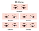
        • Strabismus is defined as any form of ocular misalignment. The cover/uncover test is used to diagnose strabismus
        • There are many types of strabismus, which are defined by the direction of misalignment
          • Exotropia: out-turning of eyes
          • Esotropia: in-turning of eyes (see ReelDx video in presentation tab)
          • Hypertropia: Upward deviation of eyes
          • Hypotropia: Downward deviation of eyes
        • Treatment options can include glasses, occlusion therapy, orthoptic exercises, surgery, or a combination of these therapies
    1. Metallic foreign bodies may leave a rust ringirrigation and removal with sterile swab. Intraocular foreign bodies require immediate surgical removal by an ophthalmologist
      1. Cerumen impaction—buildup obstructs the auditory canal and is the most common cause of conductive hearing loss
        • Abnormal Rinne test—bone conduction is better than air conduction
        • Weber—sound lateralizes to the affected side (tuning fork is perceived more loudly in the ear with a conductive hearing loss)
        TX: Irrigation after several days of softening with carbamide peroxide (Debrox) or triethanolamine (Cerumenex)
      2. Edema with cheesy white discharge, palpation of the targus is painful, Pseudomonas aeruginosa (swimmer’s ear), S. aureus (digital trauma), malignant otitis externa is commonly seen in diabetics
      3. External Ear Trauma

        Trauma to the external ear may result in hematoma, laceration, avulsion, or fracture
        • Subperichondrial hematoma
          • Blunt trauma to the pinna may cause a subperichondrial hematoma and accumulation of large amounts of blood between the perichondrium and cartilage
          • This can interrupt the blood supply to the cartilage and render all or part of the pinna a shapeless, reddish purple mass
          • Avascular necrosis of the cartilage may follow
          • The resultant destruction causes the characteristic cauliflower ear of wrestlers and boxers
          • The most appropriate course of action for this patient is to refer immediately for I & D by an ENT specialist for the best results
          • The cartilage of the pinna requires vascular supply from the perichondrium. If deprived of blood, the devascularized tissue can become permanently damaged, resulting in the so-called “cauliflower ear”
      1. Benign tumor of the Schwann cells (the cells which produce myelin sheath) – most commonly affects the vestibular division of the 8'th cranial nerve. Slowly progressive unilateral hearing loss, tinnitus, disequilibrium. Diagnose with MRI treat with surgery or stereotactic radiation therapy
      2. Barotrauma presents with ear pain and hearing loss that persists past the inciting event, associated with pressure changes. Common injury in divers or while flying, sudden onset of pain that may resolve with a "pop."
      3. Ear fullness, popping of ears, underwater feeling, intermittent sharp ear pain, fluctuating conductive hearing loss, tinnitus. All children < 7 years old have some ET dysfunction (based on the angle of the eustachian tube) will resolve with age
      4. Acute onset, vertigo + hearing loss, tinnitus of several days to a week. Usually, viral absence of neurologic deficits. Diazepam or meclizine vertigo, promethazine for nausea
        • Central vertigo: more gradual onset and vertical nystagmus. Unlike peripheral vertigo, it does not present with auditory symptoms. Romberg Sign. Brainstem vascular disease, arteriovenous malformations, tumors, multiple sclerosis, and vertebrobasilar migraine.
        • Peripheral (inner ear) sudden onset, nausea/vomiting, tinnitus, hearing loss, and horizontal nystagmus. Labyrinthitis, benign paroxysmal positional vertigo, endolymphatic hydrops (Ménière syndrome), vestibular neuritis, and head injury.
      1. Painless otorrhea, brown/yellow discharge with strong odor, caused by chronic eustachian tube dysfunction which results in chronic negative pressure and inverts part of the TM causing granulation tissue that over time, erodes the ossicles and leads to conductive hearing loss. Surgical removal.
      2. Age 2 and under, limited mobility of the TM with pneumotoscopy. S. pneumoniae 25%, H. influenzae 20%, M. catarrhalis 10%, first line Amoxicillin, second line Augmentin, macrolides if pen allergic, complications mastoiditis and bullous myringitis.
      3. Pain, otorrhea, and hearing loss/reduction, most heal spontaneously, keep clean and dry, treat with antibiotics, the only class of antibiotics that are non-ototoxic are Floxin drops.Surgery if persists past 2 months.
    1. The most common causes of hearing impairment/loss are cerumen impaction, eustachian tube dysfunction (secondary to upper respiratory tract infection [URI]), and increasing age (presbycusis)
      • Weber testTuning fork is placed on center of the head and see if sound lateralizes - Sound lateralizes to affected ear in conductive hearing loss, Sound lateralizes to unaffected ear in sensorineural hearing loss
      • Rinne test: Tuning fork placed on mastoid and then up to the ear (should continue to hear) conductive hearing loss if bone > air, sensorineural hearing loss if air > bone
      1. Complication of acute otitis media. Fever, otalgia, pain & erythema posterior to the ear, and forward displacement of the external ear. Organisms: S. pneumoniae, H. influenzae, M. catarrhalis, S. aureus, S. pyogenes. IV antibiotics (ceftriaxone), drainage of middle ear fluid.
      2. Vertigo attacks lasting hours, classic triad of low-frequency hearing loss, tinnitus with aural (ear) fullness and vertigo, treat with low salt diet, diuretics (HCTZ + triamterene) to reduce aural pressure
      3. Perceived sensation of sound in the absence of an external acoustic stimulus; often described as a ringing, hissing, buzzing, or whooshing. 90% is associated with sensorineural hearing loss – caused by loud noise, presbycusis, medications (aspirin, antibiotics, aminoglycosides, loop diuretics and CCBs), Meniere's disease, acoustic neuroma.
    2. Insects must be immobilized prior to removal. Drown insects with mineral oil or viscous lidocaine before attempting removal, After irrigation, if the child is uncomfortable, consider treating with topical pain agents such as benzocaine-antipyrine
    • Thyroid neoplastic disease (blueprint endocrinology): Papillary is the most common type
    • Oral Cancer: Most often squamous cell carcinoma (blueprint dermatology) - It has been estimated that the use of tobacco and alcohol account for up to 80 percent of cases of squamous cell carcinoma of the head and neck
    • Branchial Cleft Cyst: Cyst appearing after URI anterior to sternocleidomastoid, most common lateral neck mass
    • Thyroglossal duct cyst: Hyoid or sub-hyoid soft mass that rises with tongue protrusion - most common midline neck mass
    • Lymphadenopathy: Unilateral, painless, persistent, and cervical-think lymphoma (blueprint hematology)
    • Leukoplakia: A white oral lesion that is painless and cannot be rubbed or scraped off. Lesions are often linked with tobacco, alcohol, or denture use. About 5% are dysplastic or squamous cell carcinomas. If there is an associated erythematous appearance (erythroplakia), there is a higher risk of dysplasia or cancer (90%)
    1. Kiesselbach's Plexus or Little's Area is the most common site for anterior bleedssphenopalatine artery (Woodruff’s plexus) is generally the source of severe posterior nosebleed, direct pressure for 15 minutes, posterior balloon packing is used to treat posterior epistaxis
    2. Teardrop-shaped growths that form in the nose or sinuses, usually benign, associated with allergic rhinitis. Samter's triad: Asthma, Aspirin sensitivity, and nasal polyps. Consider Cystic Fibrosis when multiple polyps are seen
    3. Clear nasal drainage, pruritus, pale, bluish, boggy mucosa, allergic shiners, IgE mediated mast cell histamine release, intranasal decongestants not to be used more than 3-5 days may cause rhinitis medicamentosa
    4. After URI. Sinus pain/pressure (worse with bending down and leaning forward). Facial tap elicits pain. Viral: Most common, symptoms < 7 days. Bacterial: Symptoms 7+ days and associated with bilateral purulent nasal discharge. Indications for antibiotics in rhinosinusitis include duration of symptoms >10 days without improvement, Augmentin 875 BID, kids Amoxicillin x 10-14 days. Organisms: S. pneumoniae, H. influenzae, M.catarhalis. Chronic: Plainview X-ray (waters view) is a good initial screening, CT is the gold standard.
    1. Angioedema

      • Gingivitis: the patient should be counseled about increased risk for cardiovascular events.
      • Gingival Hyperplasia: Overgrowing of gums so that it blocks the teeth, commonly caused by medications: phenytoin, CCBs, and cyclosporine.
      • Vincent's Angina: “Trench Mouth” necrotizing gingivitis: characterized by the “punched-out” appearance of the gingival papillae.
      • Dental abscessPoor dental health is a risk factor for dental abscess or facial cellulitis. Treat with IM ceftriaxone and amoxicillin.
      1. Single or multiple small, shallow ulcers with yellow-gray fibrinoid center with red halos, biopsy should be considered for ulcers lasting more than 3 weeks, viscous lidocaine 2–5% applied to ulcer QID after meals until healed
      2. Immunocompromised, young patients.  Painful, white fluffy patches that can be scraped off and may bleed when scraped (candidiasis can "come off"), leaving an erythematous, friable base. Diagnose with Potassium Hydroxide (KOH) prep
      3. Deep neck space infections most commonly arise from a septic focus of the mandibular teeth, tonsils, parotid gland, deep cervical lymph nodes, middle ear, or sinuses
        • Classic manifestations of these infections include high fever, systemic toxicity, and local signs of erythema, edema, and fluctuance
        • Computed tomography (CT) is the imaging modality of choice for the diagnosis of deep neck space infection
        • The most common organisms isolated from deep neck space infections are viridans streptococci
        TX: Antibiotics, aspiration or surgical drainage should be performed
      4. Unvaccinated patient leaning forward, drooling, stridor and distress (tripod position, muffled voice), H. influenza type B (Hib). Lateral radiograph: Thumbprint sign. Secure airway, IV Ceftriaxone, and IV fluids.
      5. HSV type 1, vesicular lesions all in the same stage of development, prodromal period of tingling discomfort or itching
      6. Almost always viral, hoarseness following a URI, consider squamous cell carcinoma if hoarseness persists > 2 weeks, history of ETOH and or smoking, laryngoscopy is required for symptoms persisting > 3 wks.
      7. Acute pharyngitis (ReelDx + Lecture)

        • Viral - Adenovirus most common
        • Mononucleosis: Epstein Barr virus, fever, sore throat, lymphadenopathy, splenomegaly, atypical lymphocytes, + heterophile agglutination test (monospot). Symptomatic and avoid contact sports, antibiotics such as amoxicillin or ampicillin may cause a rash.
        • Consider gonorrhea pharyngitis in patients with recent sexual encounters, or with non-resolving pharyngitis
        • Fungal causes in patients using inhaled steroids
        • Group A Streptococcal pharyngitis: S. pyogenes. Centor Criteria: Absence of cough, Exudates, Fever, Cervical lymphadenopathy. Throat culture is gold standard, Penicillin is first line, Azithromycin if Pen allergic. Complications: Rheumatic fever and post-strep glomerulonephritis.
      1. Sialadenitis is a bacterial infection of a salivary gland (S. aureus) usually caused by sialolithiasis (obstructing stone) in the salivary gland.  Acute swelling of the cheek, which worsens with meals. Diagnose with CT, ultrasonography, or MRI.
      2. Mumps is caused by Paramyxovirus (Mumps), in adult males look for an associated orchitis
    2. Avulsed teeth
      • Avulsed permanent teeth should be reimplanted immediately by the first capable person (eg, the injured child, a parent, teacher, coach, or primary care provider)
      • Remove debris by gentle rinsing with saline or tap water; do not attempt to sterilize or scrub the tooth
      • The tooth should be placed back in the socket ideally within 15 minutes and up to one hour (or longer if stored in cold milk)
      • Avulsed primary teeth should not be reimplanted because of the potential for injury to the developing tooth bud
      1. Temporomandibular Disorders (TD)

      2. Smokers, alcohol abuse. Painless, precancerous (SCC) white lesions on the side of the tongue that cannot be scraped off

St. Patty's Day Sale! Get 20% off your SMARTY PANCE purchase (Two days only)

X