 Lecture


50 y/o with acute onset syncope and weakness
Patient will present as → a 30-year-old obese white female presents with fatigue and generalized weakness for several weeks. Physical exam reveals pale nail beds, spoon nails, mucosal pallor, and an atrophic tongue. Upon further questioning, the patient reveals a "craving for ice and inanimate objects." Laboratory data shows a microcytic, hypochromic appearance to the RBCs, an elevated TIBC, low serum iron of 16 µg/dl, and low plasma ferritin of 12 µg/dl.

To watch this and all of Joe Gilboy PA-C's video lessons you must be a member. Members can log in here or join now.
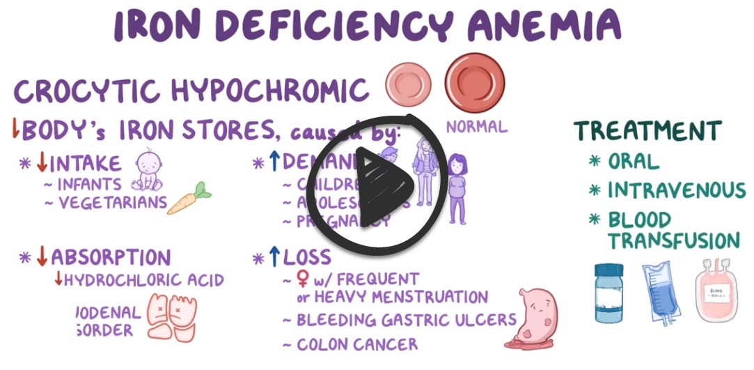
↓ MCV (microcytic), ↑ TIBC, ↓ Ferritin (low iron stores) ↓ MCH (hypochromic)
Iron deficiency is the most common cause of anemia and usually results from blood loss
- In men, the most frequent cause is chronic occult bleeding, usually from the GI tract.
- In premenopausal women, menstrual blood loss is a common cause
- Another possible cause of blood loss in men and women is chronic intravascular hemolysis
- Fatigue with exercise, palpitations, shortness of breath, weakness, headaches, and tinnitus
- Tachycardia, tachypnea on exertion, pallor, glossitis, angular cheilitis, pica, koilonychia, jaundice, and splenomegaly
RBCs tend to be microcytic and hypochromic, and iron stores are low as shown by low serum ferritin and low serum iron levels with high serum total iron binding capacity

"Lead poisoning is a common question on the PANCE/PANRE look for Basophilic stippling and remember treatment is with EDTA."
Other causes of ↓ MCV include lead poisoning (look for this in a patient with neurological symptoms), sideroblastic anemia, basophilic stippling, and thalassemia. Treatment is with EDTA.
CBC Findings
- Hemoglobin/Hematocrit (Hgb/Hct):
- <13.5 g/dL or Hct <39% (men)
- <12 g/dL or Hct <37% (women)
- <2× standard deviation of normal for age
- Mean Corpuscular Volume (MCV): ↓ (microcytic)
- Red Cell Distribution Width (RDW): ↑ (anisocytosis)
- Reticulocyte Count: ↓ (due to impaired erythropoiesis)
Iron Studies
- Serum Iron: ↓ (commonly <30 mcg/dL)
- Ferritin: ↓ (<15 ng/mL is diagnostic)
- Total Iron Binding Capacity (TIBC): ↑
- Transferrin Saturation: ↓
Peripheral Blood Smear
- Poikilocytosis with pencil or cigar-shaped RBCs
- Microcytosis and hypochromia
Additional Workup
- Hemoccult testing: If GI bleeding is suspected
- Bone marrow biopsy: Rarely needed unless etiology is unclear
Screening Recommendations
- Routine Hgb/Hct screening:
- 12 and 18 months (infants)
- 12 years (females)
- Ferrous sulfate 3 mg/kg once or twice daily (20% elemental iron) – Give between meals with juice (not milk)
- Ferrous fumarate (33% elemental iron) 100-200 mg/day in 2-3 doses
- Ferrous gluconate (12% elemental iron) 3-6 mg/kg/day in 3 doses
Side Effects
- Liquid preparations—gray staining of teeth or gums
- Brush teeth or rinse with water after administration
- GI upset (ferrous gluconate better tolerated)
Pearls
- Six weeks to correct anemia
- Six months to replete iron stores
- Recheck blood counts every 3 months x 1 year
Iron can be provided by various iron salts (eg, ferrous sulfate, gluconate, fumarate) or saccharated iron PO 30 min before meals (food or antacids may reduce absorption)
- A typical initial dose is 60 mg of elemental iron (eg, as 325 mg of ferrous sulfate) given once/day or bid
- Larger doses are largely unabsorbed but increase adverse effects especially
- Ascorbic acid either as a pill (500 mg) or as orange juice, when taken with iron, enhances iron absorption without increasing gastric distress
- The response to treatment is assessed by serial Hb measurements until normal RBC values are achieved
- Hgb rises little for 2 weeks but then rises 0.7 to 1 g/wk until near normal, at which time rate of increase tapers
- Anemia should be corrected within 2 mo
- Increasing reticulocyte count is an indication that iron is working
- A subnormal response suggests continued hemorrhage, underlying infection or cancer, insufficient iron intake, or, very rarely, malabsorption of oral iron
 Osmosis Osmosis |
|
 |
 Iron deficiency anemia accounts for more than half of anemia cases worldwide. It is typically caused by malnutrition (decreased ingestion of meat, eggs, iron-fortified foods and leafy greens), as well as malabsorption (IBD, parasitism, celiac disease). Hemorrhage is another reason for this type of anemia, which may be caused by heavy menstruation, parasitism, malignancy or ulceration. This is a microcytic, hypochromic anemia which is caused by decreased heme synthesis. Labs typically show decreased reticulocytes, and decreased ferritin, which is an iron-storing protein. There is also an increased red cell distribution width, which helps distinguish iron deficiency anemia from thalassemia.
| Iron deficiency anemia | Play Video + Quiz |
| Microcytic anemia causes | Play Video + Quiz |
| Anemia lab values | Play Video + Quiz |
| Iron (ferrous sulfate) | Play Video + Quiz |
Question 1 |
folic acid Hint: Vitamin B12 and folate deficiency present with macrocytic cells and are treated with vitamin B12 and folate respectively. | |
vitamin B12 Hint: See A for explanation. | |
prednisone Hint: Prednisone is used to treat immune-mediated hemolytic anemias which present with normocytic, normochromic red blood cells. | |
ferrous sulfate |
Question 2 |
Iron deficiency | |
Vitamin B12 deficiency Hint: Vitamin B12 deficiency is associated with macrocytic anemia. | |
Folate deficiency Hint: Folate deficiency is associated with macrocytic anemia. | |
G6PD deficiency Hint: G6PD deficiency is not associated with a low MCV. |
Question 3 |
Iron deficiency Hint: causes of microcytic anemia. | |
Anemia of chronic disease Hint: causes of microcytic anemia. | |
Sideroblastic anemia Hint: causes of microcytic anemia. | |
Folate deficiency |
Question 4 |
Chronic gastrointestinal blood loss Hint: The most important cause of iron deficiency anemia in adults is chronic blood loss, especially gastrointestinal blood loss. | |
Celiac disease Hint: Celiac disease is an occult cause of iron deficiency through poor absorption in the gastrointestinal tract. | |
Excessive vomiting | |
Zinc deficiency Hint: Zinc deficiency is causes of poor iron absorption |
Question 5 |
Anemia of chronic disease Hint: reduced serum iron, normal or raised serum ferritin, Iron present in bone marrow. | |
Iron deficiency anemia | |
Sideroblastic anemia Hint: raised serum iron, raised serum ferritin, Iron present in bone marrow. | |
Thalassemia trait Hint: normal serum iron, normal serum ferritin, Iron present in bone marrow. |
Question 6 |
Ferritin level | |
Reticulocyte count | |
Haptoglobin level | |
Transferrin level |
Question 7 |
Can present as pica, koilonychia, cheilosis, and dysphagia. Hint: See B for explanation | |
The reticulocyte count may be high. | |
Is treated with ferrous sulfate. Hint: See B for explanation | |
Can be caused by zinc deficiency. Hint: See B for explanation |
|
List |
References: Merck Manual · UpToDate