

The NCCPA™ PANCE Dermatology Content Blueprint includes three disorders under the skin integrity category, with diabetic ulcers also addressed within the PANRE blueprint for comprehensive coverage.
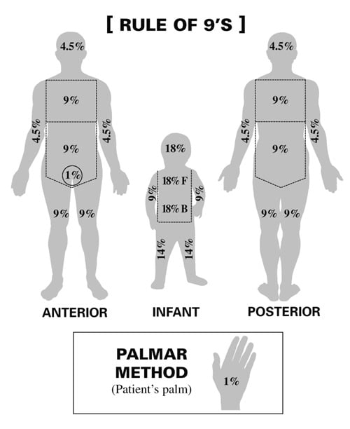
Body percentage:
- Rule of 9’s: Head 9%, Each arm 9%, Chest 9%, Abdomen 9%, Each anterior leg 9%, Each posterior leg 9%, Upper back 9%, Lower back 9%, Genitals 1%
- Palmar method: Patient’s palm equates to 1%
Depth of burn (current terms, former degree in parentheses):
- Superficial (formerly 1st degree, e.g., sunburn): Epidermis only; red, dry, blanches with pressure, tender, no blisters; heals in about a week without scarring
- Superficial partial-thickness (formerly 2nd degree): Epidermis + upper dermis; pink, blistered, moist, very painful, blanches; heals in 1 to 3 weeks with minimal scarring
- Deep partial-thickness (formerly 2nd degree): Epidermis + deep dermis; wet or waxy, mottled white to red, sluggish blanching, painful mainly to pressure; heals in more than 3 weeks with scarring and usually needs grafting
- Full thickness (formerly 3rd degree): Entire dermis destroyed; tough, white/leathery or charred, painless, no blanching; requires excision and skin grafting
- Fourth-degree (deep burn): Extends beyond the skin into fascia, muscle, tendon, and/or bone (classic with high-voltage electrical injury); never heals without surgery


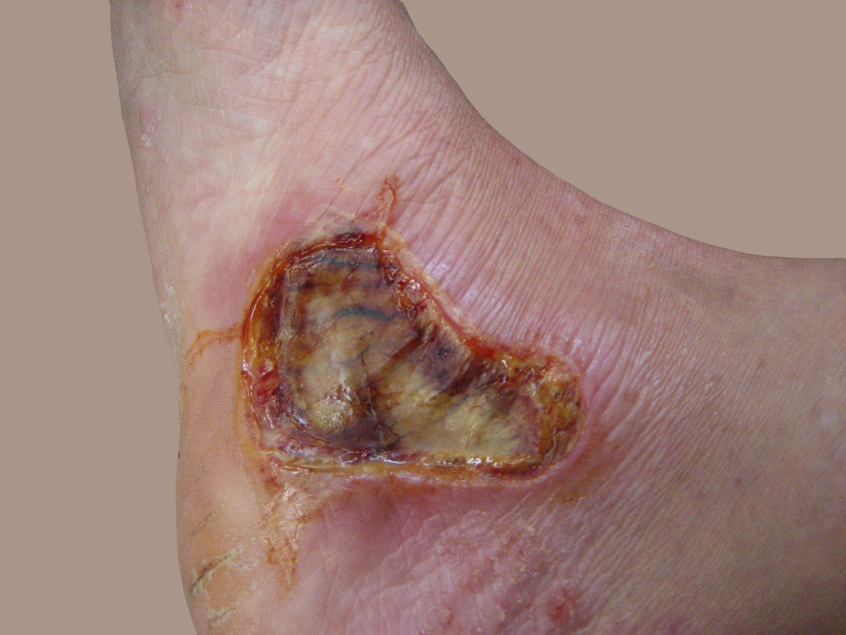

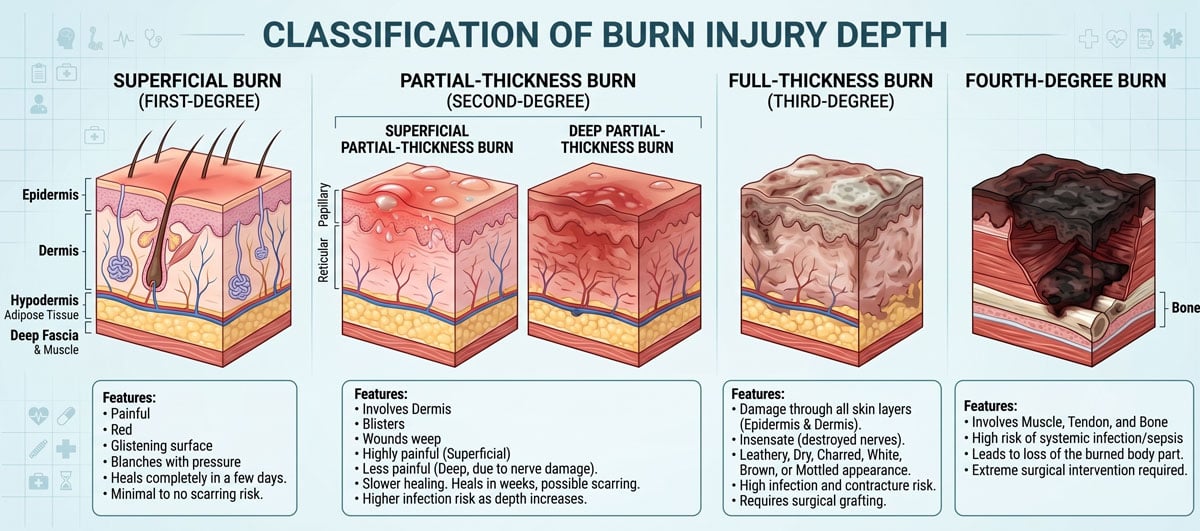 Burn depth: superficial (formerly first-degree), partial-thickness (formerly second-degree, superficial vs deep), full-thickness (formerly third-degree), and fourth-degree. Click to enlarge.
Treatment: Monitor ABCs, fluid replacement, and sulfadiazine. Children with > 10% total body surface area and adults with > 15% total body surface area burns need formal fluid resuscitation
Diabetic ulcers are chronic, non-healing wounds, typically on the feet, caused by neuropathy, poor circulation, and impaired immunity in diabetic patients
- Plantar surface, metatarsal heads, heels, and pressure points are common sites
- Risk factors include peripheral neuropathy, PAD, foot deformities, poor footwear, prior ulcers, and poor glycemic control
- Often painless, with surrounding callus, erythema, swelling; may show infection (purulence, odor) or signs of osteomyelitis
- DX: Clinical exam, probe-to-bone, monofilament testing, cultures if infected, X-ray/MRI for osteomyelitis, ABI (may need toe pressures if falsely elevated)
- TX:
- Wound care: Debridement, moist dressings
- Offloading: Total contact cast or protective footwear
- Antibiotics: Empiric, then culture-directed
- Glycemic control: Essential for healing
- Revascularization: If PAD is severe
- Surgery: For deep infection, gangrene, or nonresponse
- Complications: Osteomyelitis, cellulitis, gangrene, amputation
- Prevention: Daily foot checks, podiatric care, proper shoes, tight glucose control

Suture removal — The timing of suture removal varies with the anatomic site
- Eyelids – Three days
- Neck – Three to four days
- Face – Five days
- Scalp – 7 to 14 days
- Trunk and upper extremities – Seven days
- Lower extremities – 8 to 10 days
Follow-up visits — Most clean wounds do not need to be seen prior to removal unless signs of infection develop. Highly contaminated wounds should be seen for follow-up in 48 to 72 hours. It is imperative that clear discharge instructions are given to every patient regarding signs of wound infection.
| Suture material | Knot security | Wound tensile strength | Tissue reactivity | Workability | Anatomic site |
| Nylon (Ethilon) | Good | Good | Minimal | Good | Skin closure anywhere |
| Polybutester (Novafil) | Good | Good | Minimal | Good | Skin closure anywhere |
| Polypropylene (Prolene) | Least | Best | Least | Fair | Skin closure anywhere. Blue dyed suture useful in dark-skinned individuals. |
| Silk | Best | Least | Most | Best | Rarely used |
| Suture material | Knot security | Wound tensile strength | Security (days)* | Tissue reactivity | Anatomic site |
| Fast-absorbing gut | Poor | Least | 4 to 6 | Most | Face |
| Vicryl Rapide | Good | Fair | 5 to 7 | Minimal | Face, scalp, undercast/splint |
| Surgical gut | Poor | Fair | 5 to 7 | Most | Face (rarely used) |
| Poliglecaprone 25 (Monocryl) | Good | Fair | 7 to 10 | Minimal | Face, consider in contaminated wounds needing deep closure |
| Chromic gut | Fair | Fair | 10 to 14 | Most | Mouth, tongue, nailbed |
| Polyglactin (Vicryl) | Good | Good | 30 | Minimal | Deep closure, nailbed, mouth |
| Polyglycolic acid (Dexon) | Best | Good | 30 | Minimal | Deep closure |
| Polydioxanone (PDS) | Fair | Best | 45 to 60 | Least | Deep closure |
| Polyglyconate (Maxon) | Fair | Best | 45 to 60 | Least | Deep closure |
Most wounds should be covered with an antibiotic ointment and a nonadhesive dressing immediately after laceration repair
- The dressing should be left in place for 24 hours, after which time most wounds can be opened to air.
Prophylactic antibiotics may decrease the risk of infection in some animal and human bites, intraoral lacerations, open fractures, and wounds that extend into cartilage, joints, or tendons
- Allow animal bites, especially in non-cosmetic areas such as the hand and foot are to heal by secondary intention

The sacrum and hip are most often affected. Reposition every 2 hours
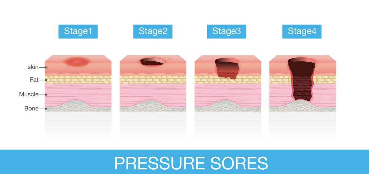
- Stage 1: erythema of localized area, usually non-blanching over the bony surface
- Stage 2: partial loss of dermal layer, resulting in pink ulceration
- Stage 3: full dermal loss, often exposing subcutaneous tissue and fat
- Stage 4: full-thickness ulceration exposing bone, tendon, or muscle. Osteomyelitis may be present
Wound management by stage of the ulcer
- Stage I: aggressive preventive measures, thin-film dressings for protection
- Stage II: occlusive dressing to maintain healing, transparent films, hydrocolloids
- Stages III-IV: Debridement of necrotic tissue. Exudative ulcers will benefit from absorptive dressings such as calcium alginates, foams, and hydrofibers. Dry ulcers require occlusive dressing to maintain moisture, including hydrocolloids, and hydrogels.
Risk factors: age >65, impaired circulation, immobilization, undernutrition, incontinence
DX: based on observation and staged according to classification
TX: debridement ⇒ depends on the extent of necrosis; surgical closure may be necessary; vacuum-assisted closure uses negative pressure to reduce wound edema and remove debris/reduce bacterial load
© logo3in1 by Adobe Stock