
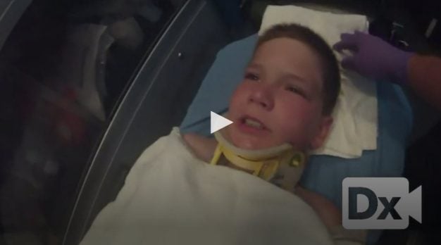
11 y/o male with back pain following a diving injury
Patient will present as → a 33-year-old female involved in a motor vehicle accident in which she is rear-ended in slow-moving traffic at less than 10 MPH. She presents to the ER complaining of localized neck stiffness and pain. On physical exam, she has paraspinal tenderness in the cervical region. She has limited motion secondary to pain. Her motor, sensory, and reflex exam are normal in her upper and lower extremities. Radiographs are obtained and show normal cervical spine AP and lateral radiographs.
All trauma patients have a cervical spine injury until proven otherwise
- Cervical clearance can be performed with a physical exam and radiographically
- CT to bottom of first thoracic vertebra replacing conventional radiographs as initial imaging in most trauma centers
- Removal of cervical collar WITHOUT radiographic studies is allowed if the patient is awake, alert, and not intoxicated AND
- Has no neck pain, tenderness, or neurologic deficits AND has no distracting injuries
Fractures at the level of the spinal cord (above L1/2) are much more vulnerable to neurologic injury than injuries below and require a more urgent treatment
Thoracic spine (T2-T10)
- Fractures from T2-T10 are rare due to increased stability of the thoracic spine
- Is a vascular watershed area so vascular injury can lead to cord ischemia
- Burst fracture
- Osteoporotic compression fracture
- Fracture dislocation (rare but leads to paralysis in 80%)
Thoracolumbar region (T11 to L2)
- More commonly affected by spine trauma due to fulcrum of motion (intersection between the stiff thoracic spine and increased motion of lumbar spine)
- More than 50% of all thoracic and lumbar fractures occur in this region
Lumbar region (L3 to S1)
- The spinal cord ends and cauda equina begins at the level of L1/L2
- Injuries below L1 have a better prognosis because the nerve roots (cauda equina and nerve roots within thecal sac) are affected as opposed to the spinal cord
Magerl classification (of thoracic spine injuries)
- Type A
- Compression caused by axial loading
- Type B
- B1: ligamentous distraction injury posterior
- B2: osteoligamentous distraction injury posterior
- Type C
- Multi-directional injuries, often fracture dislocations
- Very unstable with high likelihood of neurologic injury
- Multi-directional injuries, often fracture dislocations
AO classification (of thoracolumbar spinal fracture)
- Type A: Compression injuries
- Type B: Distraction injuries
- Type C: Torsional injury
| Level | Patient Function |
| C1-C3 | - Ventilator dependent with limited talking. - Electric wheelchair with head or chin control |
| C3-C4 | - Initially ventilator dependent, but can become independent - Electric wheelchair with head or chin control |
| C5 | - Ventilator independent - Has biceps, deltoid, and can flex elbow, but lacks wrist extension and supination needed to feed oneself - Independent ADL’s; electric wheelchair with hand control, minimal manual wheelchair function |
| C6 | - C6 has much better function than C5 due to ability to bring hand to mouth and feed oneself (wrist extension and supination intact) - Independent living; manual wheelchair with sliding board transfers, can drive a car with manual controls |
| C7 | - Improved triceps strength - Daily use of a manual wheelchair with independent transfers |
| C8-T1 | - Improved hand and finger strength and dexterity - Fully independent transfers |
| T2-T6 | - Normal UE function - Improved trunk control - Wheelchair-dependent |
| T7-T12 | - Increased abdominal muscle control - Able to perform unsupported seated activities; with extensive bracing walking may be possible |
| L1-L5 | - Variable LE and B/B function - Assist devices and bracing may be needed |
| S1-S5 | - Various return of B/B and sexual function - Walking with minimal or no assistance |
Obtain radiographs of the entire spine (concomitant spine fractures in 20%)
- CT scan indications
- Fracture on plain film
- A neurologic deficit in the lower extremity
- Inadequate plain films
- MRI is useful to evaluate for
- Injury to anterior and posterior ligament complex
- Spinal cord compression by disk or osseous material
- Cord edema or hemorrhage
Most thoracic and thoracolumbar fractures (burst and compression) can be treated nonoperatively when the patient is neurologically intact
- Treat in orthosis for 6 to 12 weeks depending on the degree of instability
- Indications for surgery include progressive neurologic deficits
- Myelomalacia seen on MRI
- Gross spinal instability
Question 1 |
Anterior cord syndrome Hint: Anterior cord syndrome presents with paraplegia or quadriplegia, loss of lateral spinothalamic function with preservation of posterior column function. | |
Central cord syndrome | |
Brown-Séquard syndrome Hint: Brown-Séquard syndrome is a condition associated with hemisection or damage to one half of the spinal cord, resulting in a loss of sensations like pain, temperature, touch, as well as paralysis or loss of muscle function in some parts of the body. | |
Complete cord transection Hint: Complete cord transection would affect motor and sensory function distal to the lesion. | |
Cauda equina syndrome Hint: Cauda equina syndrome typically presents as low back pain with radiculopathy. |
References: Merck Manual · UpToDate