Chest/rib disorders (PEARLS)
The NCCPA™ Musculoskeletal Content Blueprint chest and rib disorders (PEARLS)
Q
Flashcards ready when you areYou have opened several Quizlet sets recently. Loading this one only when needed helps prevent a temporary Quizlet limit.
 Osmosis Osmosis |
 |
| Chest/rib Deformities |
The most common is pectus excavatum (sunken chest or funnel chest) or pectus carinatum (pigeon chest)
- Less common types of chest wall abnormalities include Poland's syndrome, Jeune's syndrome, and defects of the ribs and sternum
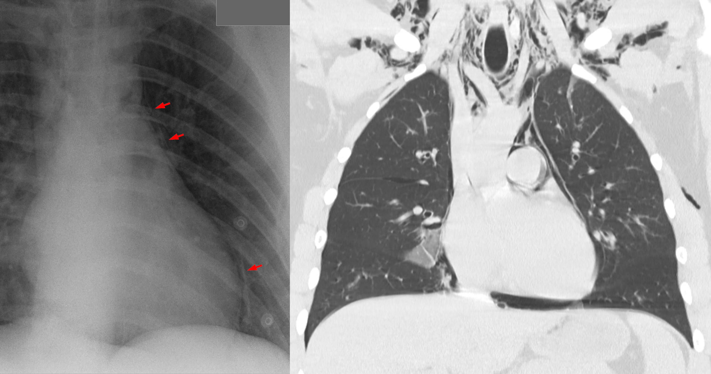
- Suspected from visual examination of the anterior chest and chest x-rays and chest CT are useful in the diagnosis
Treatment includes orthotic bracing, physical exercise, and surgery
 Pectus excavatum in a teenager.
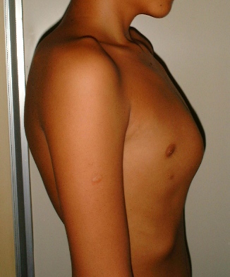 Pectus carinatum in a teenager. |
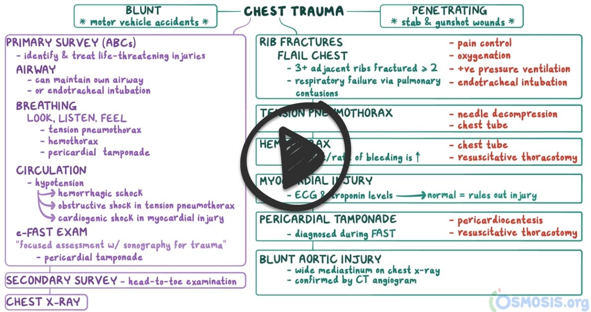
| Chest/rib Trauma |
Thoracic trauma can be distinguished by the mechanism of injury
Blunt trauma refers to mechanisms causing increased intrathoracic pressure such as car collisions (the most common cause of thoracic trauma) and falls
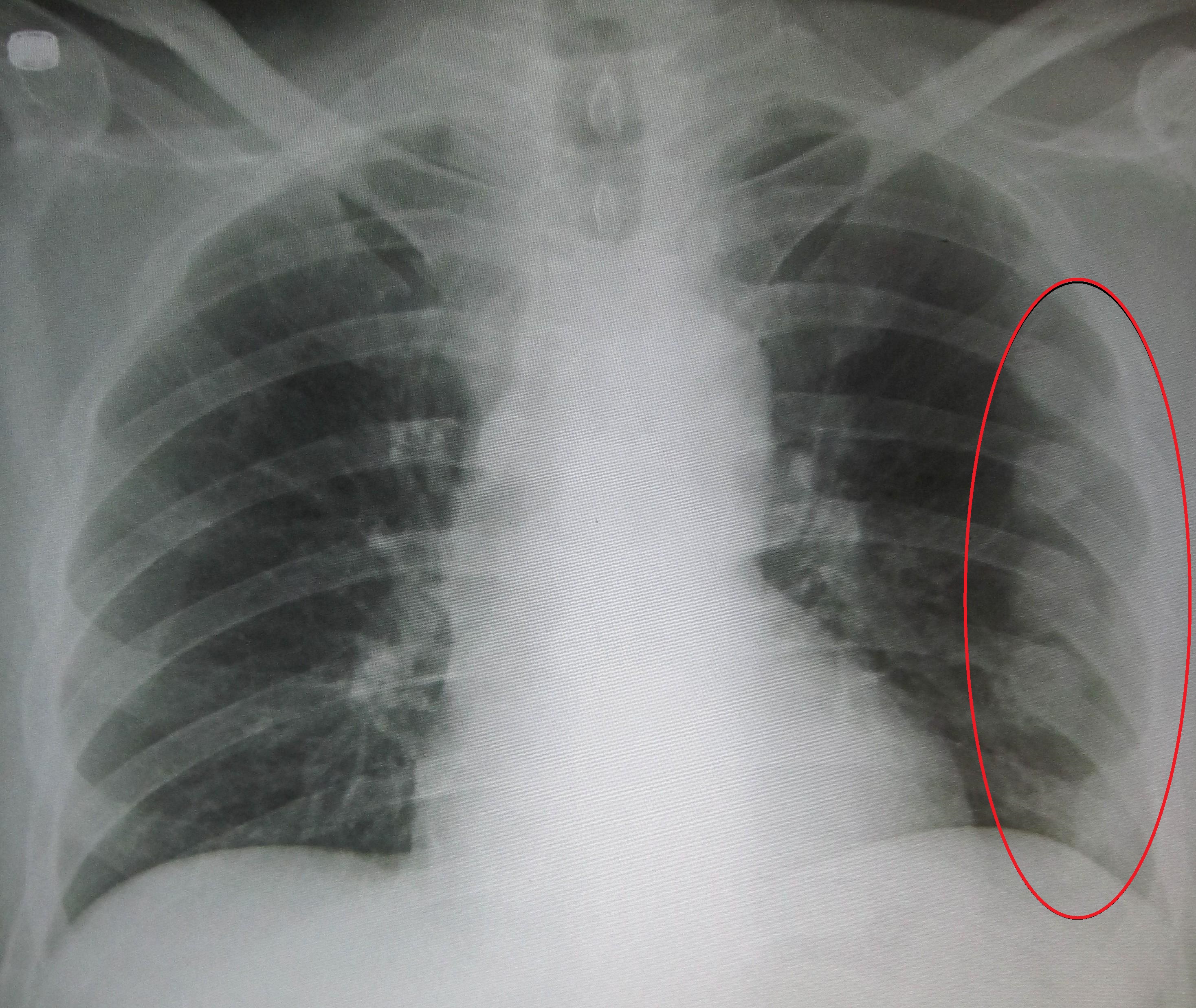
- Rib fractures are the most common blunt thoracic injuries. Ribs 4-10 are most frequently involved. Patients usually report inspiratory chest pain and discomfort over the fractured rib or ribs. Physical findings include local tenderness and crepitus over the site of the fracture.
- Flail chest involves three or more consecutive rib fractures in two or more places, which produce a free-floating, unstable segment of chest wall
- Sternal fractures cause pain, tenderness, bruising, and swelling over the fracture site. CPR has also been known to cause thoracic injury
- Pulmonary contusion resulting from blunt trauma to the chest causing interstitial edema that impairs compliance and gas exchange
- Ruptured diaphragm - should be considered in patients who sustain a blow to the abdomen and present with dyspnea or respiratory distress
- Blunt myocardial injury - dysrhythmias, ST changes
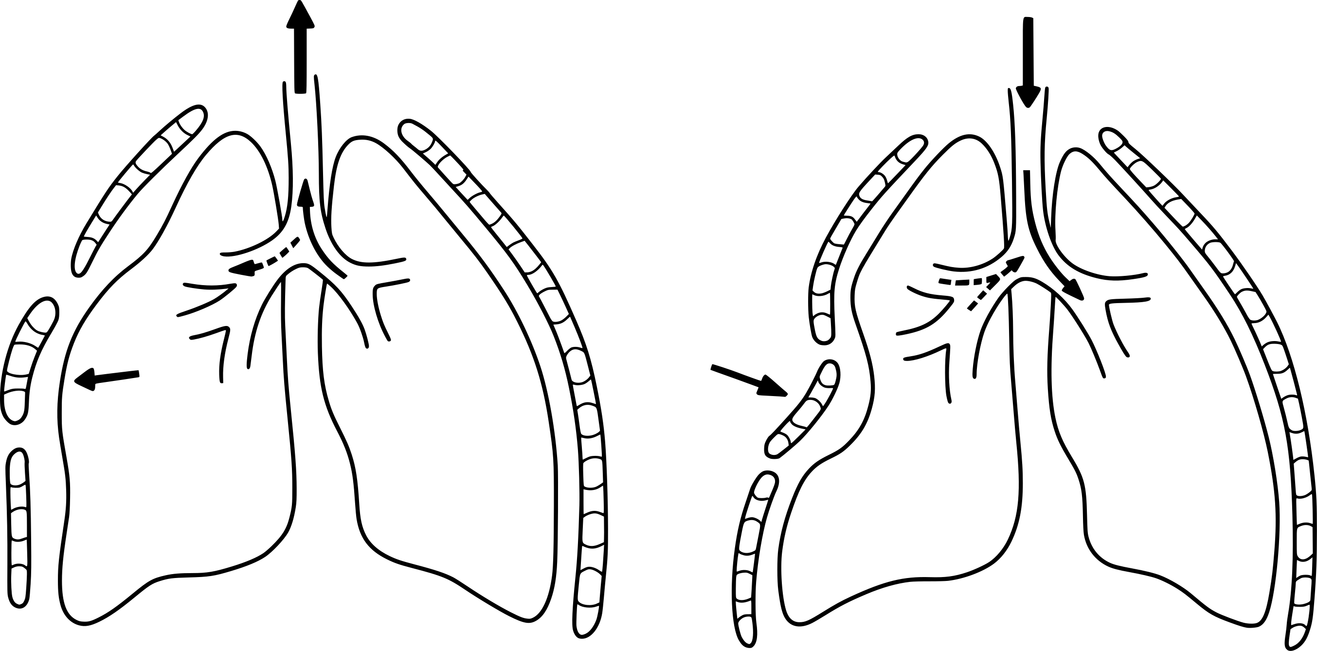
- Pneumothoraces in blunt thoracic trauma are most frequently caused when a fractured rib penetrates the lung parenchyma
Penetrating trauma largely refers to gunshot and stab wounds, occasionally impalement
Symptoms:
- Pain in the chest that gets worse when laughing, coughing, or sneezing, deep inspiration and overhead activities
- Difficulty breathing
- Tenderness, bruising, and swelling
Diagnosis:
Chest radiography remains the basis for initiating other investigations
- CT, aortography, bronchoscopy/endoscopy
- Hemoglobin or hematocrit values and arterial blood gas determinations offer the most useful information for treating these patients
- ECG
- Bone scan may be indicated when x-rays are negative and clinical suspicion remains
Treatment involves immediate airway management: intubate early if airway compromise suspected
- Rib fractures - the majority heal uneventfully with rest, analgesia, cessation of inciting activity for ~4-6 weeks
- Needle thoracostomy - Use large bore needle at 2nd intercostal space in the midclavicular line - the best initial step in the management of tension pneumothorax
- Tube thoracostomy - chest tube at 5th intercostal space in anterior axillary line - next step in management after needle placement in tension pneumothorax
- Oxygenation - positive pressure ventilation for flail chest
- Thoracotomy indications
- > 1500 ml total blood loss
- > 200 ml/h continued drainage of blood for > 3 hours
- Surgical repair as needed
|
Back to PANCE Blueprint Musculoskeletal (8%)

