PANCE Blueprint Endocrinology (7%)

Primary endocrine malignancy (Pearls)

Thyroid neoplastic disease
Patient will present as → a 45-year-old female who comes to your clinic with complaints of a gradually enlarging lump in her neck over the past six months. She mentions occasional difficulty swallowing and a hoarse voice that comes and goes. She also reports feeling more fatigued than usual and sometimes feeling unusually warm. Medical history is significant for her mother, having undergone thyroid surgery in her 50s, though the exact details are unknown. On examination, a firm, non-tender nodule is palpated on the right lobe of the thyroid gland. The patient’s reflexes are brisk. An ultrasound of the thyroid reveals a 3 cm hypoechoic thyroid nodule with calcifications, and a thyroid uptake scan shows decreased iodine uptake of the nodule compared to surrounding tissues.

Hoarse voice, solitary cold nodule on thyroid uptake scan

  • Most often, papillary carcinoma (80%)

Diagnostic studies:

  • Ultrasound is the best initial screen, followed by a thyroid uptake scan. Usually, normal thyroid function
  • Microcalcifications, hypoechogenicity, a solid cold nodule, irregular nodule margins, chaotic intranodular vasculature, and a nodule that is more tall than wide.
  • Fine needle biopsy for definitive diagnosis (all lesions >1 cm should be biopsied)

Treatment: Surgical resection

  • Surgical resection with chemotherapy and external beam radiation is reserved for anaplastic thyroid cancer

Papillary thyroid carcinoma on CT, PET CT and ultrasonography

Papillary thyroid carcinoma on CT, PET CT, and ultrasonography

Adrenal tumors/neoplastic disease
Patient will present as → a 43-year-old female with high blood pressure unresponsive to therapy. She complains of headaches, palpitations, and sweating. She has a history of neurofibromatosis type 1, though without any neurological deficits. She has multiple café-au-lait spots on her body. The ECG demonstrates sinus tachycardia. She is found to be hypertensive to 154/121 mmHg. Her 24-hour urine metanephrines and VMA come back elevated. Her abdominal CT demonstrates an adrenal mass.

Pheochromocytoma

  • Adrenal neoplasm - catecholamine secreting adrenal tumor - secretes norepinephrine and epinephrine autonomously and intermittently
  • Presentation: Recurrent headaches, HTN, sweating, palpitations.

Diagnosis:

  • 24-hour catecholamines, including metabolites (metanephrine and vanillylmandelic acid)
  • MRI or CT of the abdomen to visualize the tumor

Treatment:

  • Resect tumor - complete adrenalectomy
  • Medical treatment preoperative: Alpha-blocker (phenoxybenzamine) preop, phentolamine (acute HTN crisis), sodium nitroprusside (acute HTN crisis), nicardipine (acute HTN crisis)
    • Pre-op nonselective alpha blockade: phenoxybenzamine or phentolamine 7-14 days followed by beta-blocker to control HTN (NO solo beta-blocker ⇒ prevent unopposed alpha constriction ⇒ life-threatening HTN)

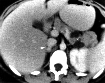
Pheochromocytoma2

CT scan demonstrating left-sided adrenal mass (pheochromocytoma)

Neoplastic syndrome (Lecture) (Prev Lesson)
(Next Lesson) Thyroid neoplastic disease (Lecture)
Back to PANCE Blueprint Endocrinology (7%)