Patient will present as → a 56-year-old male with a known history of polycythemia who suddenly complains of pain and paresthesia in the left leg. Physical examination reveals the left leg is cool to the touch, and the toes are cyanotic. The popliteal pulse is absent by palpation and Doppler. The femoral pulse is absent by palpation but weak with Doppler. The right leg and upper extremities have 2+/4+ pulses throughout.
Caused by a sudden arterial occlusion
- Remember the “six P’s” (You must know these!): Pain, Paralysis, Pallor, Paresthesia, Polar (some say Poikilothermia—you pick), and Pulselessness
- Atrial fibrillation and mitral stenosis are common causes of thrombus formation.
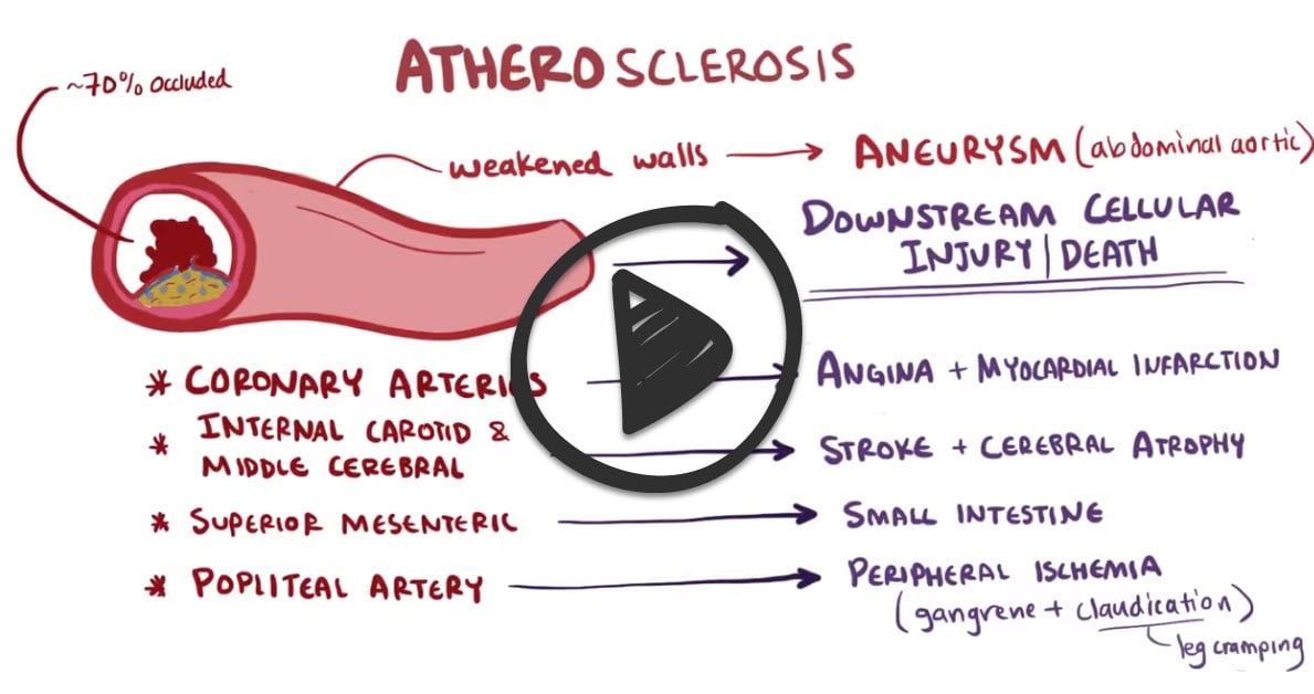
- Lower extremities are more common than upper extremities

Cyanosis of the right foot distal to an occlusion caused by acute arterial thrombosis of the right leg
Angiography is considered the gold standard for diagnosis
- ECG (looking for MI, AFib)
- Echocardiogram (+/- ) looking for clot, MI, valve vegetation
Anticoagulate with IV heparin (bolus followed by constant infusion)
- If not limb-threatening then call the vascular surgeon for angioplasty, graft, or endarterectomy
- See general surgery rotation arterial embolism and thrombosis
Flashcards ready when you areYou have opened several Quizlet sets recently. Loading this one only when needed helps prevent a temporary Quizlet limit.
 Osmosis Osmosis |
|
 |
Question 1 |
A 64 year-old patient with known history of type 1 diabetes mellitus for 50 years has developed pain radiating from the right buttock to the calf. Patient states that the pain is made worse with walking and climbing stairs. Based upon this history which of the following would be the most appropriate test to order?
Venogram Hint: See B for explanation. | |
Arterial duplex scanning | |
X-ray of the right hip and L/S spine Hint: See B for explanation. | |
Venous Doppler ultrasound Hint: See B for explanation. |
Question 1 Explanation:
Given the patient's long history of type 1 diabetes mellitus the patient most likely has vascular occlusive disease. Evaluation of arterial blood flow is assessed using the duplex scanner. X-ray of the L/S spine and right hip while not harmful may give information regarding bony structures. Venous Doppler ultrasound will not give information of arterial perfusion.
Question 2 |
A 56 year-old male with a known history of polycythemia suddenly complains of pain and paresthesia in the left leg. Physical examination reveals the left leg is cool to the touch and the toes are cyanotic. The popliteal pulse is absent by palpation and Doppler. The femoral pulse is absent by palpation but weak with Doppler. The right leg and upper extremities has 2+/4+ pulses throughout. Given these findings what is the most likely diagnosis?
Venous thrombosis Hint: See B for explanation. | |
Arterial thrombosis | |
Thromboangiitis obliterans Hint: See B for explanation. | |
Thrombophlebitis Hint: See B for explanation. |
Question 2 Explanation:
Arterial thrombosis has occurred and is evidenced by the loss of the popliteal and dorsalis pedis pulse. This is a surgical emergency. Venous occlusion and thrombophlebitis do not result in loss of arterial pulse.
Question 3 |
A patient presents with an acutely painful and cold left leg. Distal pulses are absent. Leg is cyanotic. There are no signs of gangrene or other open lesions. Symptoms occurred one hour ago. Which of the following treatments is most appropriate?
Vena cava filter Hint: Vena cava filters are used in the management of venous thromboembolic disease when anticoagulation cannot be done. | |
Embolectomy | |
Amputation Hint: Amputation is done only when no viable tissue is present. Cutting off a viable limb is never a good idea. | |
Aspirin Hint: Aspirin is used in the prevention and treatment of coronary disease and has no role in the treatment of
peripheral arterial embolism. |
Question 3 Explanation:
Embolectomy within 4 to 6 hours is the treatment of choice.
There are 3 questions to complete.
|
List |
References: Merck Manual · UpToDate