| Acid/base disorders |
Average values "24/7 40/40"
- 24 (HCO3, base) / 7.40 (pH) / 40 (CO2, acid)
Three-Step Approach to Acid-Base Disorders
- Look at your PH (7.35-7.45 is normal)
- < 7.35 = acidosis
- > 7.45 = alkalosis
- Next look at your PCO2 is it normal low or high (35-45 normal)
- ↑ CO2 and ↓PH = respiratory acidosis
- ↓ CO2 and ↑ PH = respiratory alkalosis
- If you don’t see a change in the CO2 in relation to the PH then take a look at the HCO3
- Finally, look at the HCO3 is it normal low or high (20-26 normal)
- ↓ HCO3 and ↓PH = metabolic acidosis
- ↑ HCO3 and ↑ PH =metabolic alkalosis
|
| Bladder carcinoma |
A 60-year-old white man with painless hematuria
- Painless hematuria in a smoker, transitional cell carcinoma is the most common type
- Cystoscopy with biopsy is the gold standard for initial diagnosis
Treatments include surgery, biological therapy, and chemotherapy |
| Chronic renal failure (shunts/access) |
CKD is a progression ongoing loss of kidney function (GFR) defined as less than 60 mL/min/1.73 m² or presence of kidney damage (proteinuria - urinary albumin excretion of ≥30 mg/day or equivalent, glomerulonephritis or structural damage from polycystic kidney disease) for> 3 months
Etiologies
- Diabetes is the most common cause (30% of cases)
- HTN is responsible for 25% of cases
- Chronic GN accounts for 15% of cases
- Interstitial nephritis, polycystic kidney disease, obstructive uropathy
- Any of the causes of AKI may lead to CKD if prolonged and/or if treatment is delayed
Staging CKD is a way of quantifying its severity. CKD has been classified into 5 stages
- Stage 1: normal GFR (≥ 90 mL/min/1.73 m2) plus either persistent albuminuria or known structural or hereditary renal disease
- Stage 2: mild GFR 60 to 89 mL/min/1.73 m2
- Stage 3: moderate GFR 30 to 59 mL/min/1.73 m2
- Stage 4: severe GFR 15 to 29 mL/min/1.73 m2 (symptomatic stage)
- Stage 5: kidney failure GFR < 15 mL/min/1.73 m2
Broad waxy casts in urinary sediment are a specific finding in chronic renal failure
Treatment is aimed at slowing the progression of CKD and treating reversible causes of acute deterioration
- ACE inhibitors and ARBs: slow the progression of renal dysfunction, particularly in proteinuric patients
- Managing comorbidities: Hypertensive control (< 130/80), tight glycemic control (A1C 6.5-7.5%), cholesterol control, tobacco cessation
- Maintain Hemoglobin at 11 -12 g/dl: erythropoietin, iron supplementation and antiplatelet therapy
- Dietary management: protein restriction, calcium, and vitamin D supplements, limitation of water, sodium, potassium and phosphorus
- Pneumococcal vaccination
Need for hemodialysis or kidney transplant should be coordinated with a nephrologist
- When metabolic disarray persists despite the use of restrictions and supplements, worsening acidosis, volume overload, nausea, pericardial friction rub, neuropathy, decreased mental status, worsening nutritional status, and debilitating fatigue.
- There is no evidence showing a clear benefit of initiating hemodialysis when patients are asymptomatic as opposed to uremic.
|
| Dysuria |
|
| Edema |
Edema is a swelling caused by a collection of fluid in the spaces that surround the body's tissues and organs
- peripheral edema - the lower legs or hands
- ascites - abdomen
- Chest - pulmonary edema (lungs) and pleural effusion (space surrounding the lungs)
Most common causes include chronic venous insufficiency, also a common complication of DVT
- Lymphedema surgical removal of lymph nodes for the treatment of cancer (most commonly breast cancer) can cause swelling of a limb or limbs with thickening of the skin on the side of the surgery
- Angioedema — reactions to some medications and some inherited disorders can cause fluid to leak out of the blood vessels into surrounding tissues
- Drugs — edema can be a side effect of a variety of medications, including some oral diabetes medications, high blood pressure medications, non-prescription pain relievers (such as ibuprofen), and estrogens
- Infection - peritonitis
- Hypernatremia
- Kidney disease can cause swelling in the lower legs and around the eyes
- Heart failure can cause swelling in the legs, abdomen, and lungs (pulmonary edema), causing shortness of breath
- Cirrhosis can obstruct blood flow through the liver. People can develop pronounced swelling in the abdomen (ascites) or in the lower legs (peripheral edema)
Symptoms include:
- A sensation of “heavy legs”, itching, and pain
- Hyperpigmentation, stasis dermatitis, lipodermatosclerosis (thick, brawny skin), atrophie (ivory-colored stellate scars on the legs)
- Increased size of the abdomen (with ascites)
- Difficulty breathing (with edema in the chest)
|
| Fluid and electrolyte disorders |
Q
Flashcards ready when you areYou have opened several Quizlet sets recently. Loading this one only when needed helps prevent a temporary Quizlet limit.
- Peaked T waves - Hyperkalemia
- Flattened T waves U waves - Hypokalemia
- Long QT - Hypocalcemia
- Short QT - Hypercalcemia
- Tall T waves - Hypomagnesemia
- Prolonged PR interval widened QRS - Hypermagnesemia
- Low urine sodium and polyuria, urine osmolality of less than 250 despite hypernatremia - Diabetes insipidus
- Deficient secretion of vasopressin (ADH - anti-piss-hormone) from the posterior pituitary - Neurogenic (central) Diabetes insipidus
- Caused by kidneys that are unresponsive to normal vasopressin levels - usually inherited X-linked or from lithium or renal disease - Nephrogenic Diabetes insipidus
- Hyponatremia is defined as plasma sodium concentration less than 135 mEq/L
- Hypernatremia is defined as a plasma sodium concentration greater than 145 mEq/L
|
| Nephrolithiasis |
A 31-year-old man with right flank pain radiating into the scrotum, gross hematuria, right-sided hydronephrosis, and normal abdominal x-ray
- Colicky flank pain radiating to the groin, hematuria, CVA tenderness, and nausea and vomiting
- CT scan (spiral CT) without contrast of the abdomen and pelvis is the gold standard for diagnosis
- Urinalysis will often show microscopic hematuria
- BUN and Cr levels (for evaluation of renal function) and also calcium, uric acid, and phosphate levels
Four types:
- Calcium oxalate (80%): Most common, excess oxalate, hyperparathyroidism, radiopaque - avoid grapefruit juice (makes calcium oxalate stones worse)
- Struvite (10%): Associated with chronic UTI with Klebsiella and Proteus species, radiopaque
- Uric Acid (7%): Form in individuals with persistently acidic urine - Excess meat/alcohol, gout, radiolucent
- Cystine (1%): Rare genetic, radiolucent (young boy with kidney stones)
General measures (for all types of stones)
- Analgesia: IV morphine, parenteral NSAIDs (ketorolac)
- Vigorous fluid hydration—beneficial in all forms of nephrolithiasis
- Antibiotics—if UTI is present
- Alpha-blocker therapy (Flomax) for patients with symptomatic ureteral stones >5 mm and ≤10 mm to facilitate ureteral stone passage (usually given to most patients independent of size)
- Outpatient management is appropriate for most patients. Indications for hospital admission include:
- Pain not controlled with oral medications
- Anuria (usually in patients with one kidney)
- Renal colic plus UTI and/or fever
Stones < 5 mm will have an 80% chance of spontaneous passage
- Stones > 5 – 10 mm have a 20% chance of passage and may require elective lithotripsy – patients should be considered for early elective intervention
- Stones > 10 mm are not likely to pass spontaneously. Ureteral stent or percutaneous nephrostomy (gold standard) should be used if renal function is jeopardized. Urgent treatment with extracorporeal shock wave lithotripsy can be used for renal stones of less than 2 cm or for ureteral stones of less than 10 mm
|
| Orthostatic hypotension |
Drop of > 20 mm Hg systolic, 10 mmHg diastolic, 15 BPM increase in pulse 2-5 minutes after a change from supine to standing
- Autonomic dysfunction in DM common cause, medications, tilt table testing if autonomic dysfunction is suspected
- If associated with heart rate > 15 BPM likely related to low blood volume
|
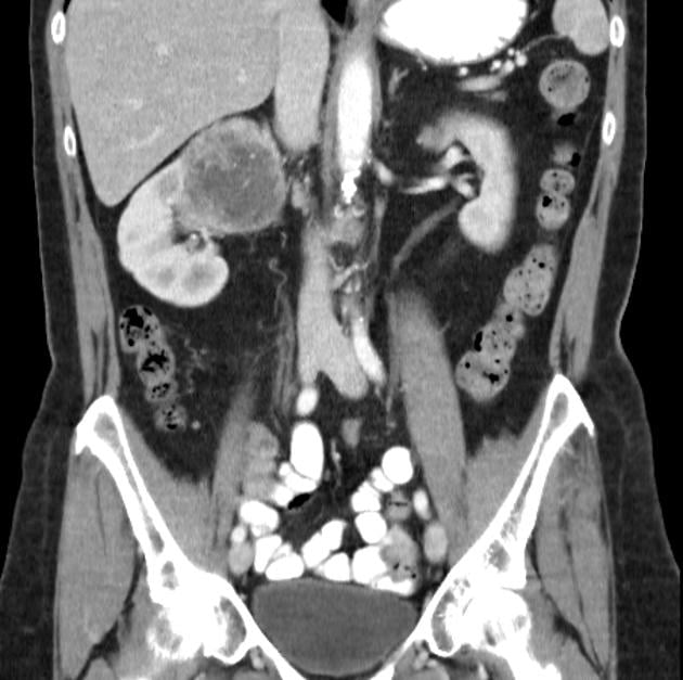
| Renal cell carcinoma |
Patient will present as → a 65-year-old man who noticed blood in his urine earlier this morning. This has never happened before and he denies any new medications or prior infection. He also reports having flank pain for the past few weeks. Medical history is significant for hypertension. He has a 40 pack-year smoking history. On physical examination, there is a firm, nontender, and homogeneous mass in the right flank. Computerized tomography (CT) scan of the abdomen is shown here. Triad of hematuria, flank pain, and abdominal mass (palpable)
- Renal clear cell = MC (80); transitional cell = second MC
- r/f: smoking
DX: ultrasound or CT then biopsy
TX: surgery with radical nephrectomy = curative |
| Renal vascular disease |
Patient will present as → a 69-year-old man with a 55-pack-year smoking history, diabetes type II, and hyperlipidemia presents to his primary care clinic for an annual exam. He has no complaints. He reports that his blood glucose has been under tight control and that he has not smoked a cigarette for the past 5 months. Vital signs are HR 69 bpm, BP 180/100 mmHg, RR 12/min, and O2 saturation 99% on room air. Physical examination is notable for bruits bilaterally just lateral of midline near his umbilicus. You initiate an anti-hypertensive medication, but his blood pressure continues to be suboptimal. Creatinine is 3.5. Narrowing of one or both of the renal arteries,
Renal artery stenosis: narrowing of one or both renal arteries most often caused by atherosclerosis or fibromuscular dysplasia
- Narrowing of artery = impeded blood flow to kidney ⇒ renovascular HTN
- Presentation: age <30 with HTN or HTN with CAD/PVD, or HTN resistant to 3+ drugs
- Patient placed on ACE who develops acute renal failure or a sharp rise in BUN/Cr ⇒ think renal artery stenosis
DX: ultrasound = first imaging in age <60
- Renal Arteriography is Gold Standard for diagnosis
- May hear a renal artery bruit on auscultation
TX: Percutaneous transluminal angioplasty (PTA) plus stent placement or with a surgical bypass of the stenotic segment |
| Testicular carcinoma |
A 22-year-old male who develops a right scrotal hydrocele with elevated serum β-HCG
- Presents as a firm, painless, non-tender testicular mass and a feeling of heaviness in the scrotum
- Seminoma is the most common type (60%)
- Risk factors include a history of cryptorchidism
- Diagnostic studies include ultrasound. Tumor markers: AFP, βHCG
Treatments include surgery, radiation, and chemotherapy |
| Urinary retention |
Postoperative Urinary Retention (POUR) is a common complication of both spinal and epidural anesthesia is a prolonged blockade of parasympathetic fibers that innervate the bladder with resultant urinary retention and the need for a urinary bladder catheter
- Obstructive causes: Urethral stricture, bladder calculi or neoplasm, foreign body
- Neurogenic causes: Multiple sclerosis, Parkinson disease, CVA, postoperative retention
- Traumatic causes: Urethral, bladder, or spinal cord injury
- Extraurinary: Fecal impaction, AAA, rectal or retroperitoneal mass
- Infectious: Local abscess, cystitis, genital herpes, zoster
Acute urinary retention
- Inability to void in the presence of a full bladder
- Risk factors: Male gender, prostatic enlargement; epidural, spinal or prolonged anesthesia; antihistamine and narcotic use; pelvic and perineal procedures M > F
- Suprapubic discomfort with urgency and inability to void
- Unable to void within 8 h after surgery or 8 h after catheter removal
- Painful
- Vomiting
- Palpable bladder on exam
- Hypotension, bradycardia, cardiac dysrhythmias
- Complication: Infection, ischemia, long-term bladder dysfunction
Chronic urinary retention
- Painless
- Develops gradually
- Frequent urination of small amounts or overflow incontinence: sensation of fullness
- Suprapubic dullness
- Rounded midline mass
Detrusor (bladder) sphincter dyssynergia
- A consequence of neurological pathology: SCI or multiple sclerosis
- Urethral sphincter muscle dyssynergically contracts during voiding causing the flow to be interrupted and bladder pressure to arise
- Obstructive cause
- Daytime and nighttime wetting
- Urinary retention
- History of UTI/Bladder infections
- Associated: Constipation and encopresis
Diagnosis:
*Physical exam (palpable bladder), bladder residual volume upon placement of a Foley catheter
Acute urinary retention
- Bladder ultrasound: 500 mL of urine
- Postvoid residual: 500 mL or greater
- Urine culture
- CBC if suspected infection
Chronic urinary retention
- Postvoid residual bladder volume by catheterization or ultrasound
- Abdominal US or CT indicated to identify suspected masses, stones, or hydronephrosis
Detrusor (bladder) sphincter dyssynergia
- Postvoid residual urine volume (PVR) >150 mL
|
| Wilms Tumor |
Child < 4 years of age with an abdominal tumor that does NOT cross the midline
Child with painless, unilateral abdominal mass with no other signs or symptoms, also known as nephroblastoma.
- HTN secondary to elevated renin levels and fever from tumor necrosis, hematuria, and anemia.
- Mean age is 3.5 years
|