The NCCPA™ Gastroenterology and Nutrition PANCE Content Blueprint covers 6 topics under the category of esophageal disorders
| Esophagitis (ReelDx) | ReelDx Virtual Rounds (Esophagitis)Patient will present as →
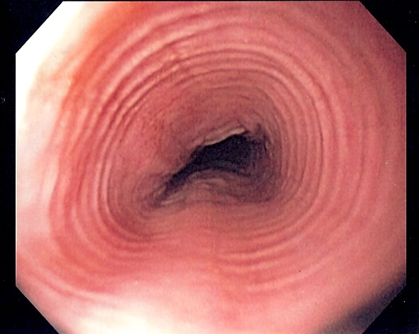
Scenario A: An immunocompromised patient on high-dose steroids presents with odynophagia (painful swallowing) and dysphagia; oral thrush is visible. This is Candida esophagitis. Scenario B: A patient on HIV therapy (especially didanosine or zalcitabine) or a patient taking a bisphosphonate without adequate water presents with sudden-onset odynophagia and substernal chest pain — pill esophagitis. Scenario C: A young atopic man has dysphagia for solids and food impaction episodes; endoscopy shows esophageal rings and “trachealization” — eosinophilic esophagitis. Esophagitis is simply inflammation that may damage the tissues of the esophagus. It can be divided into two types: 1. Non-infectious
2 Infectious - odynophagia (pain while swallowing food or liquids) is the hallmark sign This occurs mainly in patients with impaired host defenses. Primary agents include Candida albicans, herpes simplex virus, and cytomegalovirus. Symptoms are odynophagia and chest pain
DX: Upper endoscopy (EGD) with biopsy — gold standard for most types of esophagitis TX: Treat the underlying condition
 Barium swallow of the esophagus on the left side shows multiple rings associated with eosinophilic esophagitis. |
| Gastroesophageal reflux disease | Patient will present as → a 55-year-old male with complaints of heartburn, belching, and epigastric pain which is aggravated by drinking coffee, eating fatty foods, and lying down. He says it gets better when he takes antacids. GERD is the retrograde flow of gastric acid into the esophagus due to lower esophageal sphincter (LES) relaxation, causing mucosal injury and symptoms
DX: Patients with typical symptoms of GERD may be given a trial of PPI therapy. Patients who do not improve or have long-standing symptoms or symptoms of complications should be studied:
Treatment: H2 receptor blockers, proton pump inhibitors, diet modification (avoid fatty foods, coffee, alcohol, orange juice, chocolate; avoid large meals before bedtime); sleep with the trunk of body elevated; stop smoking
|
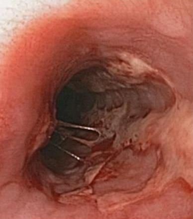
| Mallory Weiss tear | Patient will present as → a 21-year-old male with hematemesis. He is brought by his girlfriend, who reports that he and his buddies have been out drinking every night last week in celebration of his 21st birthday. He reports having vomited each night, but tonight, when he started vomiting, he noticed that there was streaking of blood. Concerned, he decided to come to the emergency department. A tear that occurs in the esophageal mucosa at the junction of the esophagus and stomach, caused by severe retching and vomiting, results in severe bleeding
DX: Diagnosed with upper endoscopy showing superficial longitudinal mucosal erosions TX: Most bleeding is self-limited (90%) and managed supportively (IV fluids, NPO, hemodynamic monitoring)
 Endoscopic image of Mallory-Weiss tear showing superficial longitudinal mucosal erosions |
| Motility disorders | Achalasia
Patient will present as → a 45-year-old patient with progressive dysphagia to both solids AND liquids, regurgitation of undigested food, chest pain, and weight loss. Symptoms worsen at night with possible aspiration/cough. Barium swallow shows “bird-beak” narrowing, and esophageal manometry confirms diagnosis. Treatment is pneumatic dilation or Heller myotomy. Achalasia is a motility disorder due to degeneration of inhibitory neurons in the Auerbach’s (myenteric) plexus → failure of LES relaxation + aperistalsis This is a FUNCTIONAL obstruction, NOT a mechanical one
DX: Barium swallow = “bird-beak” distal esophagus (classic board image finding)
TX: Pneumatic dilation: most effective non-surgical option; endoscopic balloon dilation of the LES
 Barium swallow showing dilated esophagus with retained column of barium and “bird’s beaking” suggestive of achalasia. Diffuse esophageal spasm Patient presents as → a 26-year-old male is brought to the emergency department (ED) via ambulance with a sudden onset of extreme chest pain. The patient states that he had just finished his morning run and was drinking from his water bottle when the pain began. He states that the pain was like “nothing he had experienced before” and radiated to his back, neck, and ears. He called EMS and was given 325mg aspirin, sublingual nitroglycerine, and supplemental oxygen in the field resulting in near resolution of his symptoms. In the ED, his exam is completely unremarkable except for a heart rate of 110 bpm. EKG shows sinus tachycardia, troponin and CK-MB are within normal limits, and stress test is normal. You order an upper GI contrast study which shows a corkscrew esophagus.Treatment is initiated with a calcium channel blocker (e.g., diltiazem), leading to symptom improvement. Diffuse esophageal spasm is a motility disorder of the esophagus characterized by uncoordinated, simultaneous contractions with NORMAL LES relaxation due to impaired inhibitory neural signaling.
DX: Barium swallow: classic “corkscrew” or “rosary bead” esophagus (HIGH-YIELD imaging finding)
TX: Calcium channel blockers (e.g., diltiazem) or nitrates to reduce esophageal spasm
 Corkscrew esophagus as seen in diffuse esophageal spasm Neurogenic dysphagia Patient presents as → a 32-year-old female who reports to your office complaining of nasal regurgitation with the ingestion of fluids. Sure enough, when you hand her a glass of water, and she sips the liquid, it regurgitates out her nose. You make an immediate referral to the neurologist. Three months later, when the patient returns to your office, she explains that she has been diagnosed with multiple sclerosis. Neurogenic dysphagia is a result of the faulty transmission of nerve impulses to the pharyngeal muscles, generally caused by an associated neuromuscular disease, such as myasthenia gravis, amyotrophic lateral sclerosis, MS, or stroke
Zenker diverticulum Patient presents as → a 68-year-old female seen in the emergency department with recurrent coughing spells and regurgitation after meals. Her breath is nearly unbearable upon arrival at the ED. She is also noted to have a palpable, fluctuant neck mass on physical examination. A pharyngeal pouch that develops in the proximal esophageal wall
 Outpouching of barium-filled sac as seen in Zenker diverticulum Scleroderma (Progressive Systemic Sclerosis — Esophageal Involvement) Patient presents as → a 48-year-old woman with a history of diffuse systemic sclerosis (scleroderma) who reports progressive dysphagia to solids and liquids and chronic heartburn/regurgitation. Exam shows tight, thickened skin of the hands (sclerodactyly) and telangiectasias. Workup reveals decreased LES tone and absent distal esophageal peristalsis on manometry. She is treated with proton pump inhibitors and lifestyle modification to prevent complications such as Barrett esophagus. Scleroderma esophagus is esophageal smooth muscle atrophy and fibrosis due to systemic sclerosis, causing severe hypomotility and LES incompetence
DX: Esophageal manometry (most accurate) showing absent peristalsis + low LES pressure; barium swallow may show dilated, atonic esophagus
TX: Aggressive GERD management with PPIs (first-line) + lifestyle changes (elevate head, small meals)
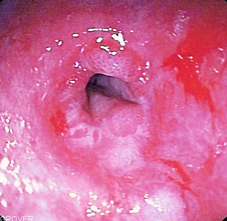
 Peptic stricture showing narrowing of the esophagus near the junction with the stomach due to chronic gastroesophageal reflux in the setting of scleroderma. Esophageal stenosis An esophageal stricture is a narrowing of the lumen of the esophagus, preventing the passage of food. Typically, it is at the distal end of the tube and is the result of scarring after chronic exposure to gastric juice due to GERD.
 Esophageal stenosis (with multiple ulcers) due to chronic reflux esophagitis |
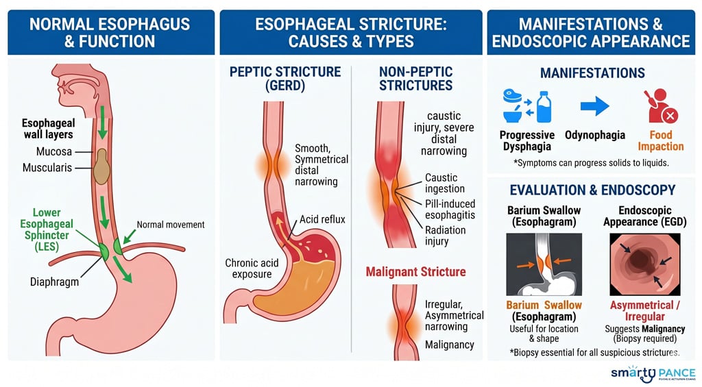
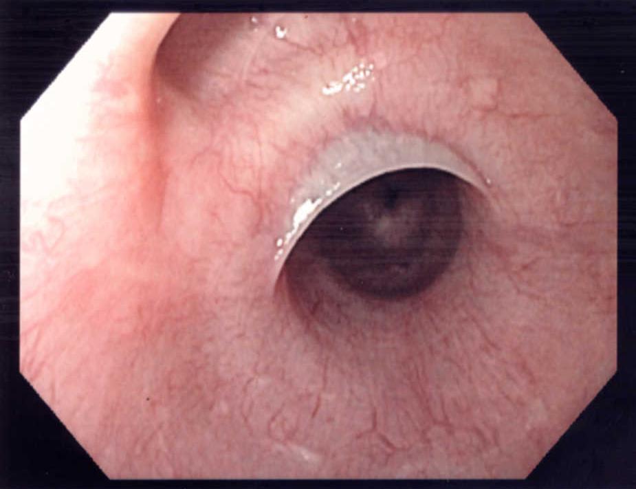
| Esophageal strictures | Patient will present as → a 60-year-old man with a 15-year history of GERD presents with progressive dysphagia to solids first, then liquids (mechanical pattern). He has had no difficulty with liquids until recently. He denies weight loss. Barium swallow shows a smooth, tapered narrowing in the distal esophagus. Upper endoscopy reveals a smooth, benign-appearing stricture (Schatzki ring / peptic stricture).
Esophageal stricture types:
Diagnosed by upper endoscopy to determine the underlying cause, exclude malignancy, and perform therapy (dilation) if needed
Treat with endoscopic dilation  Esophageal web on barium swallow: The arrowhead points to the incompletely opened upper esophageal sphincter. The arrow points to the jet phenomenon of the barium contrast when passing through the constricted area. |
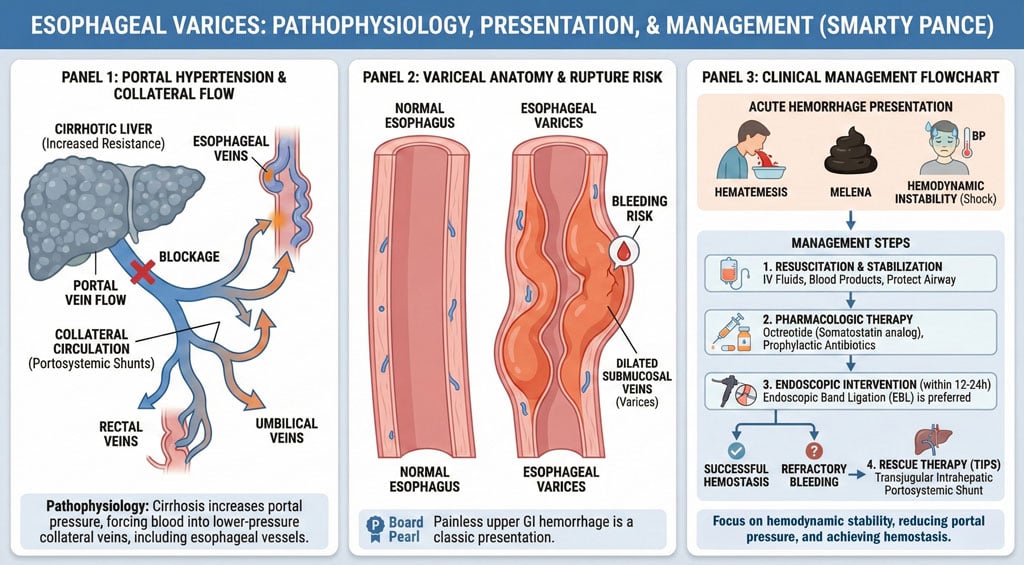
| Esophageal varices (ReelDx) | ReelDx Virtual Rounds (Esophageal varices)Patient will present as → a 64-year-old man with a history of alcoholism, tobacco use, and hypertension presents to the general surgery clinic, where he was referred for further evaluation of blood in his stool. He reports occasional abdominal pain relieved transiently with meals and one episode of painful vomiting. Recently, his stools have been black. Spider angiomas but no palmar erythema or hepatosplenomegaly are observed on the exam.
DX: Perform emergent upper GI endoscopy (once the patient is stabilized) in all patients with GI bleed ⇒ diagnostic and can be therapeutic
Screening is indicated when cirrhosis or portal hypertension is diagnosed
Treatment: Therapeutic endoscopy – endoscopic banding and IV octreotide
|
, \"Sclerotherapy\", image modification and addition of text by Stephen Pasquini PA-C, https://creativecommons.org/licenses/by- sa / 4.0 / legalcode")