

 Lecture
Patient will present as → a 37-year-old male with a history of heavy alcohol use presents to the ED complaining of rapid onset of severe mid-epigastric pain radiating to the back after eating a large meal. The pain lessens when he leans forward or lies in the fetal position. Physical exam shows low-grade fever, epigastric tenderness, diminished bowel sounds, and bruising of the flanks (Grey Turner's sign). Laboratory tests reveal elevated serum amylase and lipase, a white blood cell count of 17,000/mm³, blood glucose level of 220 mg/dL, serum LDH of 400 IU/L, and AST of 280 U/L. An abdominal CT scan shows localized dilation of the upper duodenum and a small collection of fluid in the left pleural cavity.


He is diagnosed with acute pancreatitis. Based on Ranson's criteria, his prognosis is assessed:
- On admission:
- Age over 55 years
- WBC count > 16,000/mm³
- Blood glucose > 200 mg/dL
- Serum LDH > 350 IU/L
- AST > 250 U/L
He is admitted for aggressive intravenous fluid resuscitation, pain management with opioids, and kept NPO to rest the pancreas. He is closely monitored for complications such as hypovolemia, electrolyte imbalances, and respiratory distress. Over the next 48 hours, additional parameters of Ranson's criteria are evaluated to further guide his management and prognosis. His condition gradually improves with conservative treatment, and he is eventually transitioned to a clear liquid diet before discharge.

To watch this and all of Joe-Gilboy PA-C's video lessons, you must be a member. Members can log in here or join now.
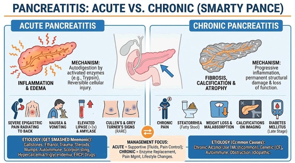
Acute Pancreatitis - epigastric abdominal pain with radiation to the back and elevated lipase - pain decreases when the patient leans forward
- It has many causes, including gallstones and chronic, heavy alcohol use
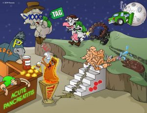
- The mnemonic GET SMASHHED is useful in recalling the most common causes:
- Gallstones, Ethanol, Trauma, Steroids, Mumps, Autoimmune disease, Scorpion sting, Hypercalcemia, Hyperlipidemia, ERCP, and Drugs
- The mnemonic GET SMASHHED is useful in recalling the most common causes:
- Cullen's sign (bruising near the umbilicus) is superficial edema and bruising in the subcutaneous fatty tissue around the umbilicus.
- Grey Turner's sign (flank bruising) refers to bruising of the flanks, the part of the body between the last rib and the top of the hip

This 40-year-old woman complained of worsening epigastric pain of five days duration. On examination, she had hypotension, a board-like abdomen, and extensive ecchymosis over her right loin (Grey Turner’s sign)

Acute pancreatitis with Cullen’s sign
Ranson’s criteria for poor prognosis
Ranson's criteria: The Ranson criteria form a clinical prediction rule for predicting the severity of acute pancreatitis. Three or more means a more severe course:
At admission:
- Age > 55
- Leukocyte: >16,000
- Glucose: >200
- LDH: >350
- AST: >250
At 48 hrs:
- Arterial PO2: <60
- HCO3: <20
- Calcium: <8.0
- BUN: Increase by 1.8+
- Hematocrit: decrease by >10%
- Fluid sequestration >6L
Chronic Pancreatitis
Clinical features are the same as those of acute pancreatitis, with the addition of fat malabsorption and steatorrhea late in the disease. Fecal fat will be elevated if malabsorption is present.
The classic triad (look for this on your exam) of pancreatic calcification, steatorrhea, and diabetes mellitus occurs in only 20% of patients
- Permanent and progressive damage to the pancreas
- Epigastric abdominal pain, weight loss, diarrhea, and pancreatic pseudocyst (a circumscribed collection of fluid rich in pancreatic enzymes, blood, and necrotic tissue)
Will have prandial epigastric pain. Labs will show increased serum lipase (more sensitive and specific than amylase, but only with elevations of threefold or greater)
- Persistent, severe epigastric pain often radiating to the back
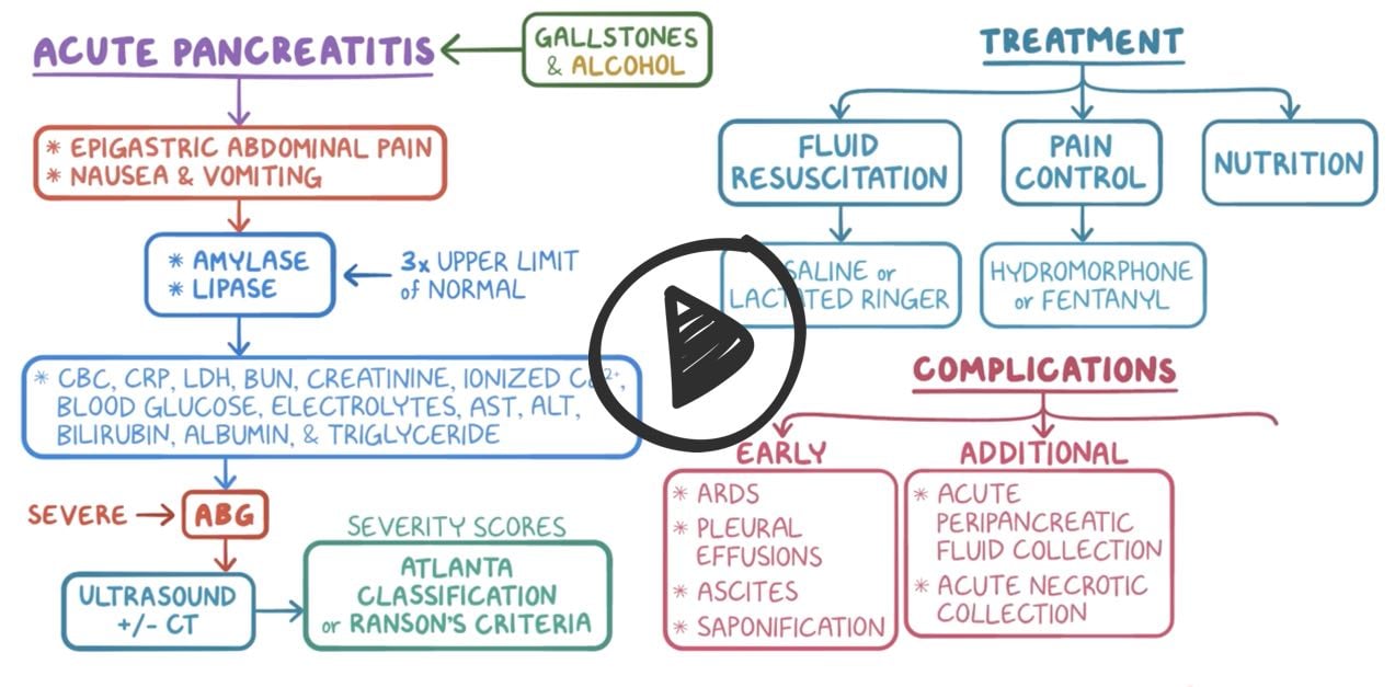
- Serum lipase or amylase ≥ 3× ULN (lipase preferred for sensitivity/specificity)
- Imaging findings consistent with pancreatitis (CT, MRI, or ultrasound)
Serum lipase is the most accurate lab test (rises within 4–8 hrs, peaks at 24 hrs, remains elevated for up to 14 days)
Amylase rises earlier but is less specific; may be normal in alcoholic or hypertriglyceridemia-induced pancreatitis
Initial lab workup should also include:
- CBC (for leukocytosis, hemoconcentration)
- CMP (for LFTs, renal function, glucose, calcium)
- Triglyceride level (if no obvious cause)
- Pregnancy test in females of childbearing age
Imaging is not required if clinical presentation and labs are diagnostic (only get imaging if diagnosis is unclear or no improvement after 48–72 hours)
- If imaging is needed:
-
- Contrast-enhanced abdominal CT is the imaging of choice—best for evaluating necrosis, complications, or unclear cases (delay ≥72 hours from onset)
- Abdominal ultrasound to evaluate for gallstones or bile duct obstruction
- MRI/MRCP is an alternative in patients with renal failure or contrast allergy
- A CT scan using a pancreatic protocol or an MRI/MRCP is the best initial diagnostic tests for chronic pancreatitis
- Sentinel loop and colon cutoff sign may be seen on plain abdominal films, but are nonspecific. Look for diminished bowel sounds as part of the exam question!

A CT scan of exudative tail pancreatitis, characterized by fluid accumulation in the fatty tissue around the pancreatic tail.
The mainstay of treatment for acute pancreatitis is supportive therapy: IV fluid resuscitation, pain control, and nutritional support
- Antibiotics for extrapancreatic infections and infected necrosis
- Endoscopic retrograde cholangiopancreatography (ERCP) for acute pancreatitis and concurrent acute cholangitis
- Complication: pancreatic pseudocyst (a circumscribed collection of fluid rich in pancreatic enzymes, blood, and necrotic tissue)
- Patients with mild pancreatitis can progress to severe pancreatitis over the initial 48 hours, often due to inadequate fluid replacement
- Referral to a tertiary center is needed if acute pancreatitis is severe or evolving/worsening

The only definitive treatment for chronic pancreatitis is to address the underlying cause, which is most commonly alcohol, low-fat diet
 Osmosis Osmosis |
|
 |
 Acute pancreatitis is an acute inflammation of the pancreas commonly caused by gallbladder disease or chronic alcohol intake. Symptoms often include abdominal pain, nausea, vomiting, anorexia, abdominal guarding and rigidity, decreased or absent bowel sounds. Elevated WBCs, generalized jaundice, and hypotension and tachycardia may also be present. Smoking is considered a risk factor.
Ranson’s Criteria
 A commonly used scaling system used to predict the prognosis and severity of acute pancreatitis, Ranson’s criteria consists of eleven parameters; five are assessed immediately on admission, and six are assessed as they develop over the next 48 hours, and the score is totaled. This card details the five admission criteria.
| Ranson’s Criteria On Admission | Play Video + Quiz |
| Ranson’s Criteria During First 48 Hours | Play Video + Quiz |
Question 1 |
Acute pancreatitis | |
Acute appendicitis Hint: Usual presentation is periumbilical pain that is later referred to the right lower quadrant. No Cullen’s sign. | |
Acute cholecystitis Hint: Presents with right upper quadrant pain that is not relieved sitting and leaning forward. Not associated with Cullen’s sign. | |
Acute gastroenteritis Hint: Presents as diarrhea with vomiting, colicky abdominal pain. Pain not relieved by sitting and leaning forward. |
Question 2 |
complete blood count with differential | |
serum amylase and lipase level | |
computed tomography (CT) scan of the abdomen with contrast
| |
comprehensive metabolic panel | |
arterial blood gases |
Question 3 |
transabdominal ultrasonography | |
contrast-enhanced CT of the abdomen | |
magnetic resonance cholangiopancreatography
| |
plain radiograph (abdominal series)
|
Question 4 |
abrupt onset of epigastric pain with radiation to the back | |
nausea and vomiting
| |
elevated serum amylase
| |
all of the above
|
Question 5 |
eliminate oral intake for the first 48 hours | |
aggressive fluid replacement
| |
calcium replacement
| |
pain control
| |
intravenous H2 receptor blockers
|
Question 6 |
a “sentinel loop” | |
the “colon cutoff sign” Hint: Gas distending the right colon that abruptly stops in the mid or left transverse colon is called the colon cutoff sign. This is caused by colonic spasm adjacent to the pancreatic inflammation but is not as common as the sentinel loop. | |
air under the diaphragm Hint: Air under the diaphragm is suggestive of a perforated peptic ulcer. | |
distention in both the small bowel and the large bowel Hint: A completely distended small and large bowel suggests a distal bowel obstruction. | |
feces throughout the colon Hint: Constipation is not associated with acute pancreatitis. |
Question 7 |
pancreatic pseudocyst formation
| |
palpable gallbladder Hint: A palpable gallbladder is seen in obstruction at the ampulla or sphincter of Oddi. | |
enlarged spleen Hint: An enlarged spleen is unlikely in this patient given the clinical history, as is an enlarged liver. | |
enlarged liver |
|
List |
References: Merck Manual · UpToDate