 Lecture
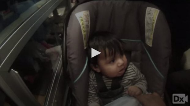
7-month-old male with dry heaves, crying, and agitation shortly after a meal
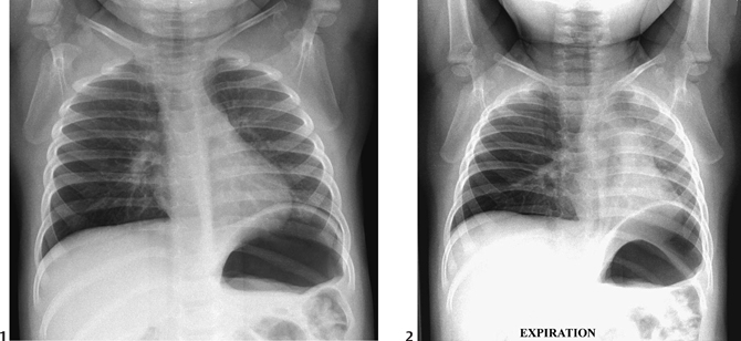
Patient will present as → a 2-year-old male child who is brought to the emergency department by his mother with a sudden onset of choking, gagging, coughing, and wheezing. Vital signs are temperature 37 ° C, pulse 120/min, and respirations 28/min. The physical examination reveals decreased breath sounds over the right lower lobe with inspiratory rhonchi and localized expiratory wheezing. The chest X-ray reveals normal inspiratory views, but expiratory views show localized hyperinflation with a mediastinal shift to the left.

To watch this and all of Joe-Gilboy PA-C's video lessons, you must be a member. Members can log in here or join now.
Aspirated solid or semi-solid object, usually lodged in the larynx or trachea
- Most often food, but can also include small toys, coins, pens, etc.
- It may be life-threatening if large enough to completely obstruct the airway
- Can lead to chronic, recurrent infection if retrieval is delayed - complications include pneumonia, acute respiratory distress syndrome, and asphyxia
Presentation depends on the location of the obstruction
- Inspiratory stridor if high in the airway
- Wheezing and decreased breath sounds if low in the airway
- 80% in the mainstem or lobar bronchus, 20% in the upper airway, right > left (most commonly lodges in the right mainstem bronchus due to more vertical anatomy)
- High-yield clue: normal child with abrupt respiratory symptoms and no fever → think aspiration, not infection
The sound of inspiratory stridor:
Risk factors include
- Institutionalization, advanced age, poor dentition, alcohol, and sedative use
All pharyngeal and airway foreign bodies are medical emergencies
CXR - expiratory radiograph may reveal regional hyperinflation of the affected side
- X-rays are normal in >50% of tracheal Foreign Body Aspirations
- X-rays are normal in >25% of Bronchial Foreign Body Aspirations
- Foreign Body Aspirations are radiolucent in >75% of Foreign Body Aspirations in ages 1 to 3 years old
ABG - necessary for appropriately evaluating ventilation, may be useful for following the progression of respiratory failure when it is of concern

Aspiration of a dental crown. This projects onto the right lower lobe bronchus.
Bronchoscopy (flexible or rigid) may help to establish the diagnosis and can also be the treatment of choice for the removal of the object
- Flexible bronchoscopy is both diagnostic and therapeutic
- Rigid bronchoscopy is preferred in children due to the wider instrument lumen (as compared to its flexible counterpart), which allows for ventilation and easier removal of objects
- Surgical removal - indicated when endoscopy is impossible or unsuccessful
- Cultures should be obtained if pneumonia is suspected
Question 1 |
stridor | |
aphonia Hint: Aphonia, inability to cough and progressive cyanosis are seen with complete obstruction of the trachea, not partial obstruction. | |
inability to cough Hint: See B for explanation. | |
progressive cyanosis Hint: See B for explanation. |
Question 2 |
Normal | |
Asthmatic Hint: Breath sounds in an asthmatic patient are usually obscured by wheezing. | |
Atelectasis Hint: Breath sounds are usually absent over an area of atelectasis. | |
Foreign body Explanations Hint: Foreign body aspiration can present with stridor, wheezing or decreased breath sounds depending on where it has lodged. |
Question 3 |
Lateral soft tissue x-ray of the neck Hint: See D for explanation. | |
Indirect laryngoscopy Hint: See D for explanation. | |
Finger sweep Hint: See D for explanation. | |
Chest x-ray |
Question 4 |
The location of an aspirated foreign body inside a patient may depend on the patient's age
| |
The likelihood of complications decreases after 24-48 hours
Hint: The likelihood of complications increases after 24-48 hours, making expeditious removal of the foreign body imperative. | |
Inflammatory changes are completely reversible
Hint: Even if the object is removed, the inflammatory changes may not be completely reversible. Some investigators believe that scar carcinoma may develop over time. | |
Foreign body aspiration is more commonly seen in females than in males Hint: The male-to-female ratio of foreign body aspiration is 2:1, depending on the study. |
Question 5 |
The most common site of esophageal impaction is at the lower esophageal sphincter (LES) at the gastroesophageal junction
Hint: Most complications of pediatric foreign body ingestion are due to esophageal impaction, usually at one of three typical locations. The most common site of esophageal impaction is at the thoracic inlet. Defined as the area between the clavicles on chest x-ray, this is the site of anatomic change from the skeletal muscle to the smooth muscle of the esophagus. The cricopharyngeus sling at C6 is also at this level and may "catch" a foreign body. About 70% of blunt foreign bodies that lodge in the esophagus do so at this location. Another 15% become lodged at the mid esophagus, in the region where the aortic arch and carina overlap the esophagus on chest x-ray. The remaining 15% become lodged at the LES at the gastroesophageal junction. | |
Most complications occur once the foreign body reaches a child's stomach
Hint: Once a swallowed foreign body reaches the stomach of a child with a normal gastrointestinal (GI) tract, it is much less likely to lead to complications | |
Migration of a foreign body from the esophagus most often leads to aortoenteric fistula
| |
Swallowed button batteries may cause substantial mucosal injury within just 2 hours |
Question 6 |
Direct examination typically provides better information than indirect laryngoscopy
Hint: In cooperative patients, indirect laryngoscopy or fiberoptic nasopharyngoscopy provides better information than a direct examination. | |
The most common cause of GI foreign bodies in adults involves accidental swallowing of small objects like toothpicks
Hint: The most common cause of GI foreign bodies in adults involves food that does not pass through the esophagus because of underlying mechanical problems. | |
In children, tracheal compression and stridor suggest a large foreign body at the upper esophageal sphincter
| |
In adults, dysphagia is associated with foreign bodies in the oropharynx but not in the esophageal regions Hint: Dysphagia is the norm in adults with esophageal foreign bodies. If the obstruction is complete, an inability to handle secretions is common. |
Question 7 |
Radiography is the recommended imaging study in all foreign body soft tissue injuries
Hint: X-rays are most useful in detecting radiopaque foreign bodies with sensitivities above 95% with adequate penetration and multiple views (anteroposterior and lateral). However, for the detection of nonradiopaque foreign bodies (eg, wood, rubber, plastic, and other plant-based foreign bodies), the sensitivity of radiography is low. | |
Fluoroscopy allows for real-time visualization and allows precise location of the foreign body using skin markers
| |
Ultrasonography use is generally discouraged in foreign body soft tissue injuries
Hint: The use of bedside ultrasonography to detect and localize soft tissue foreign bodies in the emergency department (ED) is gaining in acceptance and popularity because of its ease of use, increased availability, lack of radiation exposure, safety, and sensitivity with detection of certain types of foreign bodies | |
MRI is commonly used for foreign body detection upon initial presentation and is less valuable in nonacute presentations Hint: The use of bedside ultrasonography to detect and localize soft tissue foreign bodies in the emergency department (ED) is gaining in acceptance and popularity because of its ease of use, increased availability, lack of radiation exposure, safety, and sensitivity with detection of certain types of foreign bodies |
Question 8 |
Flexible bronchoscopy is generally preferred to rigid bronchoscopy in removing tracheobronchial foreign bodies
Hint: The rigid bronchoscope has important advantages over the flexible bronchoscope. The larger diameter of the rigid bronchoscope facilitates the passage of various grasping devices, including a flexible bronchoscope. A better chance of quick, successful extraction and better capabilities of suctioning clotted blood and thick secretions are offered by the rigid bronchoscope. | |
The bougienage method should only be performed if ingestion of a blunt object by a child was witnessed within 24 hours of the procedure
| |
Foley catheter removal is indicated for patients who have foreign bodies present for longer than 72 hours
Hint: Foley catheter removal is contraindicated in patients with foreign bodies that have been present for more than 72 hours, those with a history of esophageal disease or surgery, those who are experiencing respiratory distress, and those who are uncooperative. | |
Relaxation of the LES with glucagon is recommended more than watchful waiting for foreign bodies confirmed by imaging studies to be lodged at the LES Hint: Foreign bodies lodged at the LES can be managed by relaxation of the LES, although in some studies, success rates associated with this technique are no greater than those associated with watchful waiting. |
|
List |
References: UpToDate