The NCCPA™ PANCE Pulmonary Content Blueprint covers three categories of pulmonary neoplastic disease: Benign neoplasms, malignant neoplasms, and carcinoid tumors (neuroendocrine)
Pulmonary Neoplasms: Benign, Malignant, and Carcinoid (Neuroendocrine) Tumors
| Condition | Location & Features | Treatment |
| Benign | ||
| Hamartoma | Peripheral; benign with “popcorn” calcifications on imaging | Observation; surgical removal if symptomatic |
| Pulmonary lipoma | Peripheral; rare benign tumor of adipose tissue | Observation or resection if symptomatic |
| Fibroma | Variable; connective tissue origin, usually asymptomatic | Observation |
| Papilloma | Central (airways); squamous or glandular | Surgical excision |
| Bronchial adenoma (low-grade NET) | Central or peripheral; neuroendocrine origin | Surgical resection |
| Malignant | ||
| NSCLC - Adenocarcinoma | Peripheral; most common subtype; common in nonsmokers | Surgery ± chemo/immunotherapy |
| NSCLC - Squamous cell carcinoma | Central; linked to smoking; may cavitate | Surgery ± radiation/chemo |
| NSCLC - Large cell carcinoma | Peripheral or central; aggressive, poor prognosis | Surgery ± chemo/radiation |
| Small cell lung cancer (SCLC) | Central; very aggressive, paraneoplastic syndromes common | Chemoradiation; often unresectable |
| Mesothelioma | Pleura; associated with asbestos exposure | Surgery, chemo, palliative care |
| Pulmonary metastases | Secondary tumors; Variable locations; multiple nodules, from breast, colon, kidney, melanoma, etc. | Treat primary malignancy; palliative options |
| Carcinoid Tumors | ||
| Typical carcinoid tumor | Central; slow-growing; may cause obstruction/hemoptysis | Surgical resection |
| Atypical carcinoid tumor | Central or peripheral; more aggressive | Surgery ± adjuvant therapy |
| Carcinoid syndrome (rare) | Systemic symptoms (flushing, diarrhea, wheezing) from serotonin secretion | Octreotide; surgery if resectable |
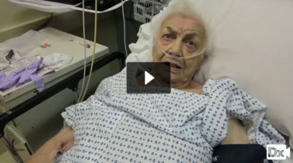
| Lung cancer (ReelDx) | Patient will present as → a 65-year-old woman with a 40-pack-year history of smoking presents with a 7 kg weight loss over the last 3 months and recent onset of streaks of blood in the sputum. PE reveals a thin, afebrile woman with clubbing of the fingers, an increased anteroposterior diameter, scattered and coarse rhonchi and wheezes over both lung fields, and distant heart sounds. ReelDx Virtual Rounds (lung cancer)Lung cancer is classified into 2 major categories
1. Non–Small Cell Lung Cancer (NSCLC) - About 85% of cases Subtypes include: adenocarcinoma, squamous cell carcinoma, large cell carcinoma, and carcinoid tumors (rare neuroendocrine tumors, not always included under NSCLC).
2. Small Cell Lung Cancer (SCLC) - About 15% of cases
DX: Chest X-ray followed by CT scan, confirmed with biopsy (bronchoscopy, CT-guided needle biopsy, or thoracoscopy)
TX: Non-small cell cancer can be treated with surgery
Small cell cancer CANNOT be treated with surgery, and will need chemotherapy Associated manifestations:
.jpg) Small cell cancer of the lung with a typical centrally located mediastinal mass (seen here on the left) |
| Carcinoid tumors | Patient will present with → a 55-year-old female who comes to the clinic complaining of recurrent episodes of flushing, particularly on her face and neck, and diarrhea for the past six months. She notes that the flushing episodes last for about five minutes and are sometimes accompanied by a feeling of warmth and palpitations. Her medical history is unremarkable except for mild asthma. On physical examination, there are no abnormal findings except for slight wheezing on lung auscultation. Given her symptoms, a 24-hour urine test for 5-HIAA is ordered, which returns elevated. An abdominal CT scan reveals a small mass in the ileum and hepatic metastases. A tumor arising from neuroendocrine cells → leading to excess secretion of serotonin, histamine, and bradykinin
DX: CT-Scan to locate the tumors
Treatment is by surgical excision and carries a good prognosis
.jpg) Carcinoid tumors are usually centrally located, arising in large airways. The arrow points to the tumor in the right hilar region. |
| Pulmonary nodules | Patient will present as → a 35-year-old female who was found to have a small (2.5 cm) pulmonary lesion on chest radiograph found incidentally after a screening exam for a positive PPD at work. The patient has no significant past medical history, and the patient is asymptomatic. < 3 cm is a nodule (coin lesion), and > 3 cm, the lesion is considered a "mass"
When managing pulmonary nodules, we follow the Fleischner Society's pulmonary nodule recommendations Steps to dealing with a pulmonary nodule:
 Chest X-ray showing a solitary pulmonary nodule (indicated by a black box) in the left upper lobe |