

 Lecture


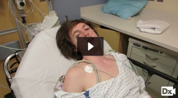
19-year-old with a history of pneumothorax and chest pain
Patient will present as → a 19-year-old male transported to the ED following a car crash. Upon arrival, he is alert and anxious and appears to be in respiratory distress. A quick assessment reveals that he sustained trauma to his face, neck, and chest. His left hemithorax appears to be expanding more than the right. He is receiving oxygen via a nonrebreathing mask. His vital signs are: respiratory rate 36 and labored. SpO2 is 85%. On physical exam, you notice decreased tactile fremitus, deviated trachea, hyperresonance, and diminished breath sounds.
Listen to his left lung field
Listen to his right lung field

To watch this and all of Joe Gilboy PA-C's video lessons you must be a member. Members can log in here or join now.
Pneumothorax is the accumulation of air in the pleural space
- Common symptoms include acute onset of ipsilateral chest pain and dyspnea with decreased tactile fremitus, deviated trachea, hyperresonance, and diminished breath sounds
Pneumothorax can be either spontaneous or traumatic
- Spontaneous (primary pneumothorax)
- Primary: occurs in the absence of underlying disease - tall, thin males between 10 and 30 years of age are at greatest risk of primary pneumothorax
- Secondary: occurs in the presence of underlying disease - asthma, COPD, cystic fibrosis, interstitial lung disease

"Spontaneous pneumothorax recurs in 30% of patients usually within 2 years. Treatment in recurrent episodes is either administration of a noxious agent into the pleural space to obliterate the pleural space or partial pleurectomy with oversewing of apical blebs or abrasion of the pleural space"
Traumatic occurs after penetrating or blunt trauma
- Tension pneumothorax can follow penetrating or blunt trauma, CPR, or positive-pressure mechanical ventilation: an injury acts as a one-way valve that lets air enter the pleural space with each breath but not leave. (An open "sucking" chest wound lets air move freely in and out; cover it with a three-sided occlusive dressing, then place a chest tube.)
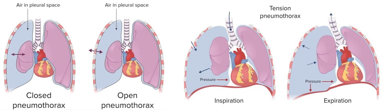
A comparison of closed, open, and tension pneumothorax (Image by Lecturio)
Pneumothorax is characterized by the acute onset of ipsilateral chest pain and dyspnea
- Physical findings depend on the size of the pneumothorax and may include unilateral chest expansion, decreased tactile fremitus, hyperresonance, and diminished breath sounds
- Tension pneumothorax is associated with a mediastinal shift to the contralateral side and impaired ventilation, leading to cardiovascular compromise
- Tension pneumothorax → penetrating injury → air in pleural space increasing and unable to escape
- Tension pneumothorax is associated with a mediastinal shift to the contralateral side and impaired ventilation, leading to cardiovascular compromise
The diagnosis of pneumothorax is a radiologic one:
- Those who are unstable should have rapid bedside imaging with pleural ultrasonography (highly sensitive and specific)
- Those with a stable presentation can wait for confirmation by upright chest radiography: a thin white visceral pleural line with no lung markings beyond it (on a supine film, look for the deep sulcus sign)
- CT for those with uncertain dx, suspected to have a loculated pneumothorax, or stable trauma patients who require CT to assess the extent of other injuries
- ABG analysis, if done, reveals hypoxemia

"A standard upright inspiratory chest X-ray is all you need: expiratory films do not detect more pneumothoraces and are no longer recommended. If the film is unclear (for example, a bulla or a skin fold could be mimicking a pneumothorax), CT is the most accurate test; bedside ultrasound (absent lung sliding, lung point) is fastest in the unstable patient."

Chest X-ray showing the features of pneumothorax
Management:
- Treatment depends on symptoms and stability, not size alone
- Minimally symptomatic, stable primary spontaneous pneumothorax → observation (conservative care) is reasonable even when the pneumothorax is large, with a follow-up CXR
- Symptomatic (significant dyspnea or hypoxemia) → needle aspiration or a small-bore chest drain first-line; an ambulatory one-way valve device is an option
- Secondary pneumothorax (underlying lung disease such as COPD) → usually admission and a chest drain
- Classic exam rule: a small pneumothorax (<15% of the hemithorax, or a rim <2 cm) often resolves on its own, while a large, symptomatic one needs drainage
"If a pneumothorax is small but the patient is mildly symptomatic or unable to return for follow-up admit the patient for observation. Give supplemental oxygen only if the patient is hypoxemic (hypoxemia should prompt a search for underlying lung disease), and avoid high-flow nasal cannula and noninvasive ventilation, whose positive pressure can worsen the leak."
Tension pneumothorax is a medical emergency
- It is a clinical diagnosis (hypotension, tachycardia, distended neck veins, absent breath sounds): do not wait for imaging. Insert a long, large-bore needle to allow air to move out of the chest, at the 4th or 5th intercostal space at the anterior-to-mid axillary line in adults (the 2nd intercostal space at the midclavicular line is an accepted alternative)
- Placement of a chest tube follows the decompression
 Osmosis |
|
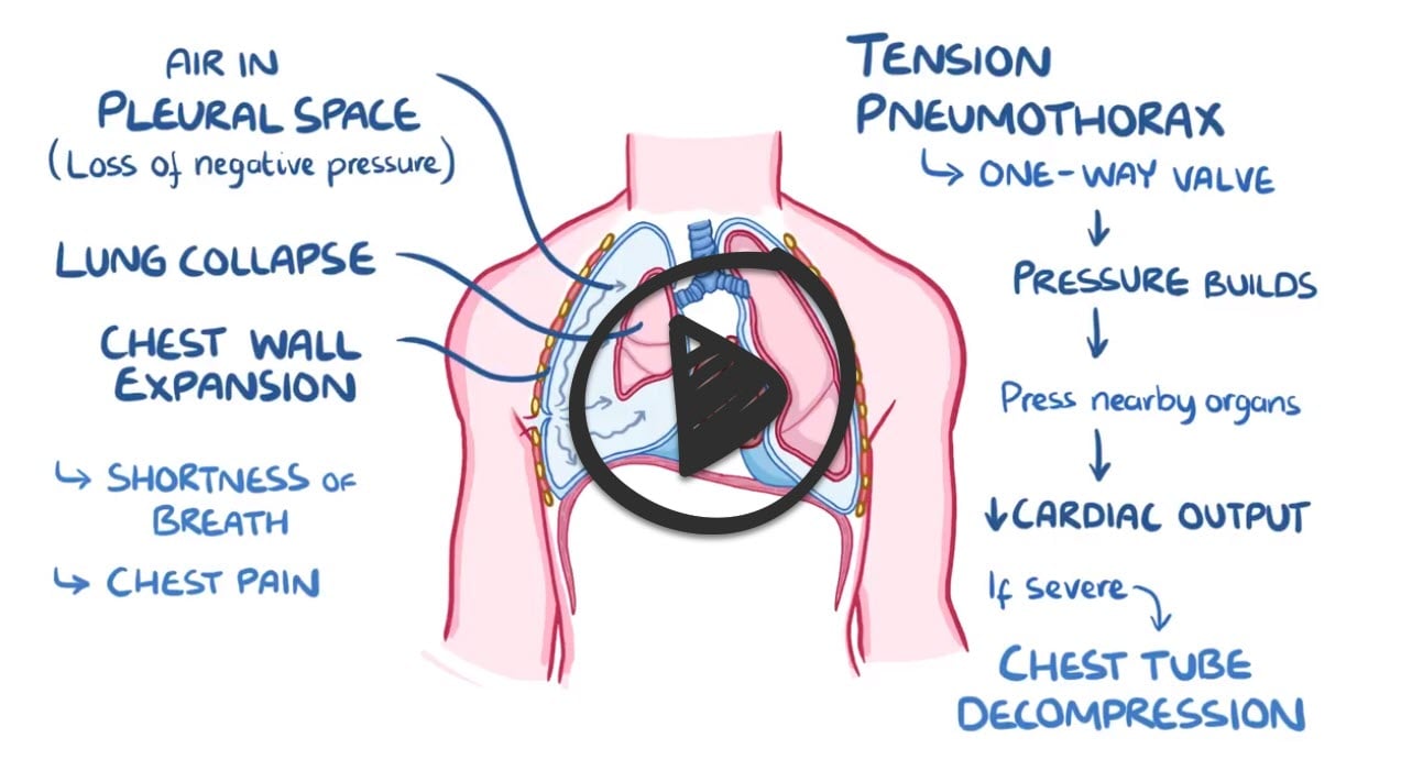 |

Tension pneumothorax is characterized by intrapleural pressure that is greater than the atmospheric pressure. It presents with sudden deterioration, hypotension, jugular venous distention, and no breath sounds. It is classically caused by trauma or positive-pressure mechanical ventilation.
Play Video + QuizSpontaneous pneumothorax

Spontaneous pneumothorax occurs due to the rupture of a subpleural apical bleb that leads to the accumulation of air in the pleural space. It is more common in tall and thin young males.
Question 1 |
Air trapping from severe asthma Hint: Asthma can cause a secondary pneumothorax, but he has no lung disease. | |
Rupture of a lung cyst from lymphangioleiomyomatosis Hint: Lymphangioleiomyomatosis causes cystic lung disease and secondary pneumothorax in young nonsmoking women, not this young man. | |
Rupture of emphysematous bullae Hint: Bullae from COPD cause secondary spontaneous pneumothorax in older smokers with known lung disease. | |
Rupture of subpleural apical blebs | |
Traumatic laceration of the visceral pleura Hint: He has no history of trauma. |
Question 2 |
Arterial blood gas Hint: An arterial blood gas may show hypoxemia or respiratory alkalosis but cannot show or exclude a pneumothorax. | |
CT pulmonary angiography Hint: CT angiography looks for pulmonary embolism; CT is reserved for an uncertain radiograph (for example, a bulla versus a pneumothorax). | |
D-dimer Hint: D-dimer helps exclude pulmonary embolism in low-risk patients; it does not diagnose pneumothorax. | |
Upright chest radiograph | |
Ventilation-perfusion scan Hint: A ventilation-perfusion scan evaluates pulmonary embolism, not pleural air. |
Question 3 |
Chest tube thoracostomy Hint: A chest tube is for unstable patients or failed observation or aspiration; in a stable, minimally symptomatic patient it adds pain, hospital days, and persistent air leaks. | |
High-flow nasal cannula oxygen Hint: Positive pressure from high-flow nasal cannula can worsen the air leak; oxygen is only needed for hypoxemia. | |
Needle decompression Hint: Needle decompression treats tension pneumothorax (hypotension, tachycardia), which he does not have. | |
Observation with a repeat chest radiograph in 4 to 6 hours | |
Thoracoscopic bleb resection with pleurodesis Hint: A definitive procedure is not routine after a first primary pneumothorax; it is reserved for recurrence, persistent air leak, or high-risk occupations. |
Question 4 |
Administer a sclerosing agent Hint: Pleurodesis prevents recurrence of pneumothorax; it has no role in the emergency treatment of tension physiology. | |
Chest radiograph Hint: Tension pneumothorax is a clinical diagnosis. Waiting for imaging in an unstable patient delays life-saving decompression. | |
Immediate needle decompression of the left chest | |
Pericardiocentesis Hint: Cardiac tamponade also causes hypotension and distended neck veins, but breath sounds are preserved and the trachea stays midline. | |
Ventilation-perfusion scan Hint: A V/Q scan evaluates for pulmonary embolism and is never appropriate in an unstable patient with signs of tension pneumothorax. |
|
List |
References: Merck Manual · UpToDate