|
Pediatric Rotation Room
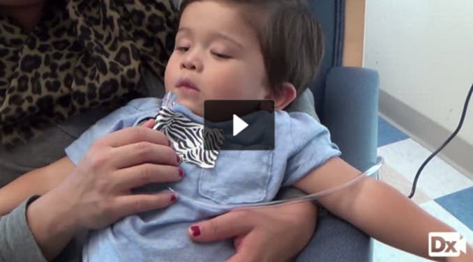 You are called to see a 2 y/o with tachypnea, hypoxemia, and fever
Patient
- Gender: Male
- Age: 2 years
- Weight: 24.5 lb/11.1 kg
- Height: 29.9 in/75.9 cm
Vitals
- Temperature: 100.1 F/37.8 C
- Blood Pressure: 110/82
- Heart Rate: 164
- Respiratory Rate: 52
- Pulse Oximetry: 86 RA
Signs and Symptoms
- 5-days fever; audible grunting; subcostal retractions; tachypnea; bilateral conjunctivitis; clear rhinorrhea; poor response to albuterol; decreased appetite; decreased urine output.
Click here to work through this patient case simulation.
Patient will present as → a 9-month-old infant with a three-day history of a mild respiratory tract infection with serous nasal discharge, fever of 38.5 C (101.4 F), and decreased appetite. Physical exam reveals a tachypneic infant with audible wheezing and a respiratory rate of 65. Flaring of the alae nasi, use of accessory muscles, and subcostal and intercostal retractions are noted. Expiratory wheezes are present. Most often caused by RSV - commonly in the fall and winter months
- Infants and young children
- Highest risk <6 months, also severe in older adults ≥75 or high-risk 50–74
- Tachypnea, respiratory distress, wheezing
- Symptoms typically peak at days 3-5 and resolve within 7-10 days
DX: Clinical DX (URI prodrome → wheezing/crackles + ↑ work of breathing); if needed → nasopharyngeal PCR; CXR NOT routine (if done: hyperinflation, peribronchial thickening) TX:
TX: Supportive care—oxygen, hydration, nasal suctioning
- Oxygen is indicated when SpO₂ persistently <90% or clinical hypoxia is present
- Hospitalization if SpO₂ <90%, apnea, moderate–severe work of breathing (retractions, nasal flaring, grunting), poor feeding/dehydration, or high-risk patient (e.g., prematurity, congenital heart disease, age <3 months)
- NO ROUTINE bronchodilators (albuterol or nebulized epinephrine), systemic steroids, or antibiotics
- Trial of albuterol may be considered in select cases (e.g., severe respiratory distress with wheezing); stop it if there is no response
- Ribavirin reserved for severe disease in immunocompromised or high-risk patients
Vaccination (view current CDC guidelines)
- Adult RSV vaccine for ≥75 and high-risk 50–74
- Pregnant individuals (32–36 weeks gestation, September–January):
- 1 dose maternal RSV vaccine → passive infant immunity
- If given ≥14 days before delivery, the infant does NOT need monoclonal antibody
- Most infants need either maternal vaccine or the monoclonal antibody (not both)
- Nirsevimab (Beyfortus) – first-line infant prevention
- All infants <8 months entering the first RSV season (unless protected by maternal vaccine)
- 8–19 months high-risk → dose before second season
- Palivizumab (Synagis) – only if nirsevimab unavailable
|
| Acute bronchitis (ReelDx) |
Patient will present as → a 23-year-old female with a one-week history of cough productive of whitish sputum. This was preceded one week prior by a URI. She denies chills, night sweats, shortness of breath, or wheezing. Temperature is 99.9°F (37.7°C). 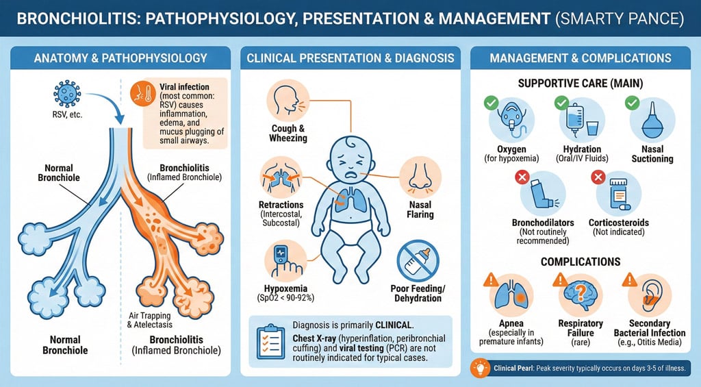 Acute bronchitis is defined by a cough >5 days; it can last 1-3 weeks Acute bronchitis is defined by a cough >5 days; it can last 1-3 weeks
Organisms:
- Most common - viral (over 90%)
- Common bacterial = M. catarrhalis
- Chronic lung patients: H. influenzae, S. pneumoniae, M. catarrhalis
Presentation:
- Cough, fever (unusual), constitutional symptoms
- Typically, less severe than pneumonia, normal vital signs, no rales, no egophony
DX: Obtain CXR if the diagnosis is uncertain or symptoms persist despite conservative treatment
TX: symptomatic and supportive - hydration, expectorant, analgesic, B2 agonist, cough suppressant
- Corticosteroids are indicated if a history of underlying reactive airway disease
- If O2 < 96% on room air, the patient should be hospitalized
- Antibiotics are indicated in the elderly, underlying cardiopulmonary disease, cough >7-10 days, or immunocompromised
- A macrolide such as azithromycin 500 mg orally once, then 250 mg orally once a day for 4 days, or clarithromycin 500 mg orally twice a day for 7 days is the preferred choice
|
| Acute epiglottitis |
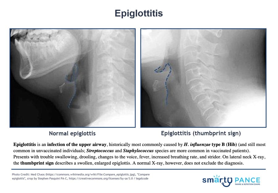
Patient will present as → a 3-year-old boy who is brought to the ER with a sudden onset of fever (104.0 F), respiratory distress, and stridor. On examination, the boy appears acutely ill. He is sitting, leaning forward with his mouth open. He has a muffled voice and is drooling. When asked, the parents report, “We don’t believe in vaccinations.” Epiglottitis is supraglottic inflammation with impending airway obstruction — a medical emergency
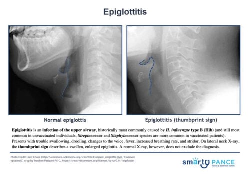
- Haemophilus influenzae type B (Hib) is the classic cause in unvaccinated children (Hib vaccine at 2, 4, 6, 12–15 mo) — but in the vaccine era most cases are Streptococcus (pneumoniae, group A), Staphylococcus aureus (including MRSA), and the condition is now more common in adults
- Key test clue: severe throat pain with minimal oropharyngeal findings + drooling/stridor = epiglottitis until proven otherwise
- vs. croup — epiglottitis is abrupt, high fever, toxic, drooling, tripod position, no cough; croup has a barking cough, hoarseness, and the steeple sign
Tripod positioning ⇒ 3 Ds of epiglottitis:
- Dysphagia
- Drooling
- Respiratory Distress
DX: High suspicion or unstable → straight to the OR — laryngoscopy is definitive ("cherry-red," swollen epiglottis) and secures the airway in the same setting
- Stable patients only: lateral neck X-ray → thumbprint sign; never send an unstable patient to radiology (or to lie flat in CT)
- Blood and epiglottic cultures after the airway is secured
TX: Airway first — anticipate intubation in a controlled setting (OR, with surgical airway backup); all patients are admitted (ICU)
- ⚠️ Keep the child calm — no tongue-depressor throat exam, no IV sticks, no supine positioning until the airway is secure — agitation can precipitate complete obstruction
- Empiric IV ceftriaxone + vancomycin (adds MRSA coverage)
- Rifampin prophylaxis for unvaccinated close contacts when Hib is the cause
|
| Croup (ReelDx) |
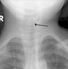
Patient will present as → a 2-year-old boy who is brought to you by his father, who is concerned about a “barking cough,” mild fever, and a hoarse voice. He reports that he had a runny nose last week that has since resolved. Physical exam reveals inspiratory stridor that is audible only when he cries; he is not drooling and swallows normally. AP neck film is shown here. Croup (viral laryngotracheitis) is a viral infection of the larynx and subglottic airway causing barking cough, inspiratory stridor, and hoarseness — peaks at 6 months–3 years, fall and early winter
- Etiology: parainfluenza virus type 1 is #1 — rhinovirus, RSV, and SARS-CoV-2 are also common causes
- Classic course: 1–3 days of coryza and low-grade fever → "seal-like" barking cough, hoarseness, and stridor that is worse at night — cough usually resolves within 3 days
- Severity is clinical (Westley score): mild = no stridor at rest; moderate–severe = stridor at rest + retractions ± agitation, lethargy, or cyanosis
- ⚠️ Drooling, dysphagia, tripod posture, toxic appearance, or high fever → NOT croup — think epiglottitis (or bacterial tracheitis if a croup-like illness suddenly worsens with high fever and toxicity)
DX: Clinical diagnosis — barking cough + stridor in a 6-month to 3-year-old; no labs or imaging needed in typical cases
- AP neck/chest film only if the diagnosis is unclear, course is atypical, foreign body is suspected, or severe croup fails to respond → "steeple sign" (subglottic narrowing) with a normal epiglottis
- Recurrent or atypical croup (outside the age range, prolonged > 7 days) → evaluate for underlying airway anomaly (subglottic stenosis, hemangioma)
TX: Single-dose dexamethasone for ALL severities — 0.6 mg/kg PO (max 16 mg); IM/IV if unable to take PO
- Mild (no stridor at rest): dexamethasone + supportive care (antipyretics, fluids) → home — humidified air/mist is unproven and is no longer a recommended therapy
- Moderate–severe (stridor at rest): nebulized racemic epinephrine + dexamethasone → observe 2–4 hours for rebound before discharge; may repeat epi every 15–20 min
- ⚠️ Admit if stridor persists after 2 epi doses, hypoxemia (SpO2 < 92%), toxic appearance, poor air entry, or age < 6 months — heliox/HFNC bridge to intubation (use an ETT 0.5–1 mm smaller) if failing
- No antibiotics — reserve for bacterial tracheitis; avoid cough/cold meds < 2 years
 AP x-ray of the neck in a child with croup demonstrating the steeple sign (narrowing of the trachea) |
| Influenza (ReelDx) |
Patient will present as → a 5-year-old with sudden onset of fever, chills, malaise, sore throat, headache, and coryza. The child is also complaining of myalgia, especially in her back and legs. On physical exam, the patient appears lethargic, has a temperature of 102.5 F, and palpable cervical lymph nodes. Breath sounds are distant with faint end-expiratory wheezes. Influenza is a viral respiratory infection caused by orthomyxovirus resulting in fever, coryza, cough, headache, and malaise
- Three strains exist: A, B, and C
Vaccination
- The CDC recommends routine annual influenza vaccination is recommended for all persons aged ≥ 6 months who do not have contraindications
Dx: rapid antigen test in the clinic, rapid serology test more accurate
Treatment is symptomatic (for most) or with antivirals ⇒ ideally< 48 hours – Tamiflu (oseltamivir), inhaled Relenza (zanamivir), IV Rapivab (peramivir), and oral baloxavir (Xofluza)
- Zanamivir and Oseltamivir both treat influenza A and B ⇒ (think Dr. “OZ” treats the flu)
- Intravenous Peramivir and Oral Baloxovir also treat influenza A and B
- Antiviral treatment reduces the duration of illness by about 1 day and should be specifically considered for high-risk patients
- Indications for antiviral treatment: hospitalized, outpatient with severe/progressive illness, an outpatient at high risk for complications (immunocompromised, pt with chronic medical conditions, >65 yo, pregnant women / 2 weeks postpartum)
 Chest X-ray in a patient with viral pneumonia demonstrating diffuse bilateral infiltrates. |
| Pertussis (Whooping Cough) |
Patient will present as → a 4-year-old boy with a severe cough following one week of cold symptoms, including sneezing, conjunctivitis, and nocturnal cough. He presents with paroxysms of cough followed by deep inspiration and occasional post-tussive emesis. 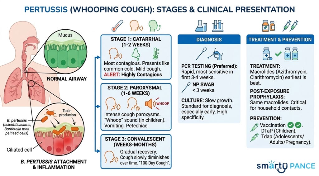 Whooping cough (pertussis) is a highly contagious respiratory tract infection marked by a severe hacking cough followed by a high-pitched intake of breath that sounds like a whoop. Whooping cough (pertussis) is a highly contagious respiratory tract infection marked by a severe hacking cough followed by a high-pitched intake of breath that sounds like a whoop.
- Gram-negative bacteria Bordetella pertussis – highly contagious
- Consider in adults with cough >2 weeks, patients < 2 years old
- Catarrhal stage: cold-like symptoms, poor feeding, and sleeping
- Paroxysmal stage: high-pitched "inspiratory whoop"
- Convalescent stage: residual cough (100 days)
Diagnosed by a nasopharyngeal swab of nasopharyngeal secretions – culture
Tx: macrolide antibiotic - preferred: azithromycin or clarithromycin
- Trimethoprim/sulfamethoxazole may be substituted in patients ≥ 2 months who cannot tolerate macrolides
- Supportive care includes hydration and dextromethorphan for cough (insufficient evidence for steroids or beta2 agonists)
- Vaccination: 5 doses – 2, 4, 6, 15-18 mo, 4- 6yrs (DTap)
- 11-18 yo = 1 dose Tdap
- Expectant mothers should get Tdap during each pregnancy, usually at 27-36 weeks
|
| Pneumonia (PEARLS) |
Presentation: Tachycardia, tachypnea, dyspnea, febrile, age 65+
Physical exam: Egophony, fremitus, rales
Chest radiograph: Infiltrates and or consolidation
Treatment:
Community-Acquired
- Adult:
- Healthy outpatients (no comorbidities):
- First line: High-dose amoxicillin
- Alternatives: Doxycycline, or a macrolide only if local pneumococcal macrolide resistance is <25% (resistance is often higher in the US)
- Outpatients with comorbidities (chronic heart, lung, liver, or kidney disease; diabetes; alcohol use disorder; cancer):
- Amoxicillin-clavulanate (or a cephalosporin such as cefpodoxime) + a macrolide or doxycycline
- OR a respiratory fluoroquinolone alone (levofloxacin or moxifloxacin)
- Inpatient (medical ward):
- Ceftriaxone + azithromycin
- OR a respiratory fluoroquinolone alone
- Child:
- First line: Amoxicillin
- Second line: 2nd or 3rd generation Cephalosporin, Clindamycin or Macrolide
Hospital-Acquired (HAC): Vancomycin + Piperacillin/Tazobactam
AIDs patients receive Bactrim prophylaxis against PJP pneumonia
CURB-65 Score for Pneumonia Severity
Estimates mortality of community-acquired pneumonia to help determine inpatient vs. outpatient treatment.
- confusion, urea >7, RR >30, Systolic BP < 90 mmHg or Diastolic BP ≤ 60 mmHg, age >65
- 0-1 = low risk, consider home tx
- 2 = probable admission vs close outpatient management
- 3-5 admission, manage as severe
| Pathogen |
Demographics |
Presentation |
| Streptococcus pneumoniae |
Most common, post-splenectomy |
Rust-colored sputum, single rigor. Lobar infiltrate |
| Mycoplasma pneumoniae |
College students, dorms |
Walking pneumonia: low temp, bullous myringitis |
| Klebsiella pneumoniae |
Alcohol abuse, chronic illness |
Currant jelly sputum |
| Legionella pneumophila |
Air conditioning, aerosolized water |
|
| Chlamydia pneumoniae |
College kids |
Long prodrome, sore throat |
| Pseudomonas spp. |
Cystic fibrosis *HAC: Ventilator-associated |
|
| Haemophilus influenzae |
COPD, smokers, post-splenectomy |
|
| Pneumocystis jiroveci |
HIV CD4 <200, immunosuppressed |
Slow onset, increased LDH, interstitial infiltrates, bilateral |
| Staphylococcus spp. |
After influenza/viral infection *HAC (MRSA) |
S. Aureus - salmon-colored/lobar |
| Fungus |
Leukemia, lymphoma, immunosuppressed, AIDs
- Histoplasma capsulatum caused by bat droppings -looks like sarcoidosis on CXR
- Cryptococcus causes meningitis
- Coccidioides (valley fever) in dry states
|
|
| Viral |
- Influenza pneumonia: Most common in adults, characterized by a more precipitous onset and fulminant course
- Adenovirus: Tends to cause symptoms fast, will present with GI symptoms and lasts about 1 week. May differentiate from bacterial mycoplasma pneumonia as mycoplasma is slow and insidious.
- RSV: Children < 1 year old
- Parainfluenza: Children 2-5 years old
|
|
|
| Respiratory syncytial virus infection (ReelDX) |
Patient will present as → a 5-month-old infant with a three-day history of a mild respiratory tract infection with serous nasal discharge, fever of 38.5 C, and decreased appetite. Physical exam reveals a tachypneic infant with audible wheezing and a respiratory rate of 65. Nasal flaring, use of accessory muscles, subcostal and intercostal retractions are noted. Expiratory wheezes and cough are present. MC cause of lower respiratory tract infection in children worldwide – virtually all get it by age 3; the leading cause of pneumonia and bronchiolitis
- Rhinorrhea, wheezing/coughing that persists for months, low-grade fever, nasal flaring/retractions, nail bed cyanosis
- Diagnosed with nasal washing, RSV antigen test; CXR can show diffuse infiltrates
Tx: Indications for hospitalization ⇒ tachypnea with feeding difficulties, visible retractions, oxygen desaturation < 95-96%
- Supportive measures include antipyretics, humidified oxygen, and nasal suctioning; systemic steroids are not routinely recommended => resolves in 5-7 days
- Bronchodilators (e.g., albuterol via nebulizer) are not recommended in bronchiolitis (per AAP)
Vaccination (view current CDC guidelines)
- Approved for individuals 60 years and older
- 1 dose of maternal RSV vaccine during weeks 32 through 36 of pregnancy, administered September through January
- To prevent severe RSV disease in infants, CDC recommends either maternal RSV vaccination or infant immunization with RSV monoclonal antibodies. Most infants will not need both
Infants and young children
- 1 dose of nirsevimab for all infants younger than 8 months born during or entering their first RSV season
- 1 dose of nirsevimab for infants and children 8–19 months old who are at increased risk for severe RSV disease and entering their second RSV season
Prophylaxis: Palivizumab (Synagis) prophylaxis, FDA-approved for children at high risk for severe RSV disease
- Premature infants born at < 29 weeks, < 1 year of age
- Premature infants born at < 32 weeks, < 1 year of age with chronic lung disease
- In the 2nd year of life for children who need supplemental oxygen or long-term corticosteroid or diuretic therapy
- Children < 24 months who have significant congenital heart disease or pulmonary hypertension
See Bronchiolitis |
| Tuberculosis (ReelDx) |
Tuberculosis
 You are called to see an 8 y/o with three months of cough, night sweats, and arthritis You are called to see an 8 y/o with three months of cough, night sweats, and arthritis
Patient
- Gender: Male
- Age: 8 years
Vitals
- Temperature: 103.6 F/39.8 C
Signs and Symptoms
- 3 months cough, night sweats; R knee edema, tenderness; slight murmur; anemic.
Click here to work through this patient case simulation.
Patient will present as → 34-year-old female nurse presents with a worsening cough of six weeks’ duration together with weight loss, fatigue, night sweats, and fever. A recent HIV test was negative. A CT scan of the chest reveals a 3 cm lesion in the upper lobe of the left lung and calcification around the left lung hilus. A sputum smear was positive for acid-fast organisms. Tuberculosis (TB) is a disease caused by bacteria called Mycobacterium tuberculosis (acid-fast bacilli)
- Presentation: fatigue, productive cough, night sweats, weight loss, post-tussive rales
- RF: endemic area, immunocompromised (HIV), recent immigrants (<5 y/o), prisoners, healthcare workers
- Transmission: inhalation of aerosolized droplets
Screening with tuberculin skin test (TST) or interferon-gamma release assays IGRAs
Mantoux Test Rules: The test is positive if induration
- > 5 mm at high risk, fibrotic changes on CXR, immunocompromised HIV/drugs, steroids/TNF antagonists daily, or close contact with pt with infectious TB
- > 10 mm in patients age < 4 or some risk factors = hospitals and other healthcare facilities, IVDU, recent immigrants from high prevalence area, renal insufficiency, prison, homeless shelter, diabetes, head/neck cancer, gastrectomy/jejunoileal bypass surgery
- > 15 mm if there are no risk factors
Diagnosis with sputum for AFB smears and Mycobacterium tuberculosis cultures – have to be 3 AFB negative
TX:
If PPD/IGRA is POSITIVE, order a CXR
- Start empiric treatment in those who likely have it
PPD positive or IGRA + CXR negative = latent TB ⇒ CDC recommends short-course, rifamycin-based, 3- or 4-month latent TB infection treatment regimens over 6- or 9-month isoniazid monotherapy
-
- Three months of once-weekly isoniazid plus rifapentine (3HP)
- Four months of daily rifampin (4R)
- Three months of daily isoniazid plus rifampin (3HR)
PPD positive or IGRA + CXR positive = active TB ⇒ Several treatment regimens are recommended in the United States for active TB disease. TB treatment can take 4, 6, or 9 months depending on the regimen.
-
- 4-month Rifapentine–moxifloxacin TB treatment regimen
- High-dose daily rifapentine (RPT) with
- Moxifloxacin (MOX): QT-prolonging agent and has been associated with cardiac arrhythmias, which may be fatal
- Isoniazid (INH) and
- Pyrazinamide (PZA)
- 6- or 9-month quad therapy (RIPE) TB treatment regimen:
-
-
- Rifampin (RIF): Orange body fluids, hepatitis - "remember R = red/orange body fluids"
- Isoniazid (INH): peripheral neuropathy (give with B6 - pyridoxine 25 to 50 mg/day)
- Pyrazinamide (PZA): Hyperuricemia (Gout)
- Ethambutol (EMB): Optic neuritis, red-green blindness - "remember E = eyes"
All are hepatotoxic, so you need to get baseline labs
- Most outpatients will be managed by your local health department to directly observe them taking TB meds for monitoring, etc.
- Patients with active TB will need two negative AFB smears and cultures in a row for therapy cessation
- Prophylaxis for household members ⇒ (example) Isoniazid for one year
- D/C therapy if transaminases > 3-5 × ULN
- Pts on INH should take supplemental Vitamin B6 (pyridoxine 25-50mg/day) to prevent neuropathy
Monitor serum creatinine; take meds on an empty stomach since food can reduce absorption, watch for hepatotoxicity, be aware of drug interactions, especially with HIV meds
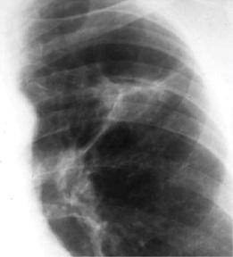
 Upper cavitary lesion on CXR associated with Tb
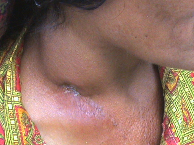 A case of long-standing tubercular lymphadenitis with sinus |