
Other pulmonary topics: high-yield conditions that are not listed under a current NCCPA Pulmonary Content Blueprint category but still appear on PANCE, PANRE and rotation exams
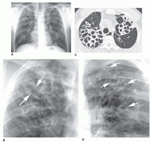
| Bronchiectasis | Patient will present as → a 25-year-old cystic fibrosis patient complaining of chronic, frequent coughing productive of yellow and green sputum. She recently recovered from a Pseudomonas spp. Pneumonia requiring hospitalization. On physical examination, you notice foul breath, purulent sputum, and hemoptysis, along with a CXR demonstrating dilated and thickened airways with “tram-track” lines and ring shadows. A condition in which the lungs' airways become dilated and damaged, leading to inadequate clearance of mucus in airways
DX: CXR = linear “tram track” lung markings, dilated and thickened airways – ring shadows (often nonspecific)
TX: daily airway clearance (cornerstone) (chest physiotherapy, hypertonic saline) + vaccines; exacerbations → 14 days of antibiotics guided by prior sputum culture (Pseudomonas → ciprofloxacin); 3 or more exacerbations per year → long-term azithromycin (rule out nontuberculous mycobacteria first); no DNase in non-cystic fibrosis bronchiectasis; surgery for localized disease, lung transplant for advanced disease 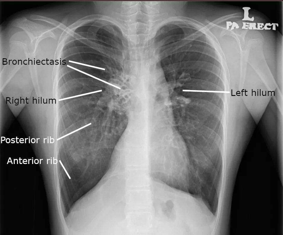 Chest X-ray of hyperinflated lung with bronchiectasis at the right upper lobe of a 12-year-old boy. Image: Cerevisae, Public domain, via Wikimedia Commons. 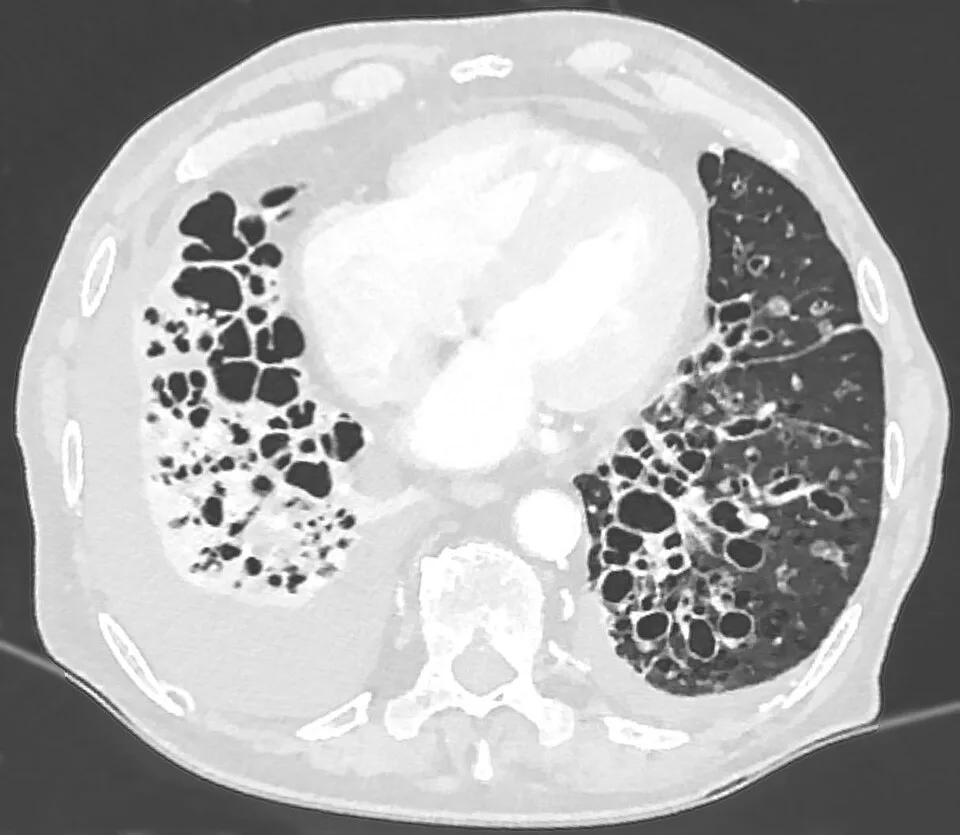 A CT scan demonstrating very pronounced bronchiectasis in a 75-year-old man, especially on the right (left in the picture). There is a florid superinfection with a large accompanying effusion. Image: Hellerhoff, CC BY-SA 3.0, via Wikimedia Commons. |