

 Lecture

Patient will present as → a 25-year-old cystic fibrosis patient complaining of chronic, frequent coughing productive of yellow and green sputum. She recently recovered from a Pseudomonas spp. Pneumonia requiring hospitalization. On physical examination, you notice foul breath, purulent sputum, and hemoptysis, along with a CXR demonstrating dilated and thickened airways with “tram-track” lines and ring shadows.

To watch this and all of Joe-Gilboy PA-C's video lessons you must be a member. Members can log in here or join now.
Bronchiectasis is permanent dilation and destruction of the bronchial walls - the common endpoint of many disorders that cause chronic airway inflammation
- A "vicious cycle": infection + impaired mucus clearance → inflammation → airway wall destruction → more mucus, bacterial colonization, and infection
Causes
- In adults, most cases are post-infectious (prior pneumonia, pertussis, tuberculosis, nontuberculous mycobacteria) or idiopathic; childhood viral infections (adenovirus, influenza, measles) are classic triggers
- Other causes: COPD, immunodeficiency (e.g., common variable immunodeficiency), allergic bronchopulmonary aspergillosis (ABPA), rheumatoid arthritis, primary ciliary dyskinesia (Kartagener syndrome - situs inversus, sinusitis, infertility), alpha-1 antitrypsin deficiency, and focal obstruction such as foreign body aspiration or tumor
- Cystic fibrosis is a classic cause (upper-lobe disease) - consider it in younger patients and test for it
Who gets it and how it presents
- More common in women and rises steeply with age
- Daily cough with copious, purulent (often foul-smelling) sputum for months to years, with recurrent chest infections, dyspnea, and fatigue
- Hemoptysis is common and usually mild (from bronchial arteries); massive hemoptysis is rare but can be life-threatening
- Crackles are common on exam; clubbing is uncommon
Organisms
- Haemophilus influenzae and Pseudomonas aeruginosa are the most commonly isolated bacteria
- Chronic P. aeruginosa infection signals more severe disease and predicts more exacerbations, admissions, and death
CXR (first test) – linear ("tram track") lung markings and ring shadows from dilated, thickened airways (often nonspecific, and may be normal)
- The diagnostic test of choice is a noncontrast high-resolution CT of the chest - airway wider than its adjacent artery (signet ring sign), thickened airway walls, lack of tapering, and airways visible near the pleura
- Distribution clues: upper lobes = cystic fibrosis; central (perihilar) = ABPA; middle lobe and lingula = nontuberculous mycobacteria
- Look for the cause: CBC, immunoglobulin levels (IgG, IgA, IgM), sputum culture (including mycobacteria), sweat chloride if cystic fibrosis is possible, and Aspergillus IgE if ABPA is possible
- Pulmonary function tests usually show an obstructive pattern

Chest X-ray of hyperinflated lung with bronchiectasis at the right upper lobe of a 12-year-old boy.

A CT scan demonstrating very pronounced bronchiectasis in a 75-year-old man, especially on the right (left in the picture). There is a florid superinfection with a large accompanying effusion
Airway clearance is the cornerstone of daily treatment
- Chest physiotherapy and airway clearance devices daily (± hypertonic saline), pulmonary rehabilitation, and vaccines (influenza, pneumococcal)
- Exacerbations → send sputum culture, then 14 days of antibiotics chosen by prior sputum cultures
- H. influenzae → amoxicillin, amoxicillin-clavulanate, or doxycycline; Pseudomonas → ciprofloxacin (IV antipseudomonal antibiotic if severe or resistant)
- 3 or more exacerbations per year → long-term macrolide (azithromycin) after ruling out nontuberculous mycobacteria; inhaled antibiotics for chronic Pseudomonas; brensocatib (oral DPP-1 inhibitor) is a newer add-on that reduces exacerbations
- Treat the cause: immunoglobulin replacement for immunodeficiency, ABPA treatment, alpha-1 antitrypsin replacement
- Avoid recombinant DNase (dornase alfa) in non-cystic fibrosis bronchiectasis (it can be harmful) and routine inhaled corticosteroids (unless asthma or COPD)
- Surgery for localized disease; lung transplant for advanced, refractory disease
 Osmosis |
|
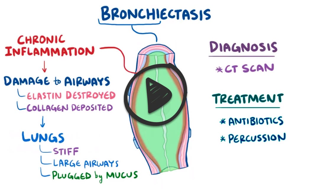 |

Bronchiectasis is permanent airway dilation caused by chronic infection and inflammation, sometimes from bronchial obstruction. The subsequent muscle and elastic tissue damage permanently dilates the airways, which may trap air in the lungs and lead to collapse of the bronchioles. Common symptoms include increased purulent sputum production and hemoptysis. In adults, most cases follow prior lung infections or are idiopathic. Poor ciliary motility from congenital causes like Kartagener’s Syndrome (primary ciliary dyskinesia) is a classic association. Other predisposing conditions are cystic fibrosis, which impairs clearance of mucus from the airways, and allergic bronchopulmonary aspergillosis (ABPA), an immune response to Aspergillus fungus.
Play Video + QuizQuestion 1 |
Allergic bronchopulmonary aspergillosis Hint: ABPA causes central (perihilar) bronchiectasis in patients with asthma or cystic fibrosis, but it is an uncommon cause overall. | |
Alpha-1 antitrypsin deficiency Hint: Alpha-1 antitrypsin deficiency is a recognized but uncommon cause; it is checked in selected patients. | |
Cystic fibrosis Hint: Cystic fibrosis is a classic cause (upper-lobe disease, often diagnosed in childhood or young adulthood), but it accounts for a minority of adult cases. | |
Postinfectious airway injury | |
Primary ciliary dyskinesia Hint: Primary ciliary dyskinesia (Kartagener syndrome) is a rare cause that usually presents in childhood with sinus and ear disease. |
Question 2 |
Bronchoscopy Hint: Bronchoscopy can help with cultures or hemoptysis but does not show the airway dilation that defines the diagnosis. | |
Noncontrast high-resolution CT of the chest | |
Pulmonary function testing Hint: Pulmonary function tests (usually an obstructive pattern) assess severity but cannot confirm bronchiectasis. | |
Sputum culture Hint: Sputum culture guides antibiotic choice but does not confirm the structural diagnosis. | |
Ventilation-perfusion scan Hint: A V/Q scan evaluates pulmonary embolism, not airway dilation. |
Question 3 |
Amoxicillin for 5 days Hint: Amoxicillin has no activity against Pseudomonas, and 5 days is too short for a bronchiectasis exacerbation. | |
Azithromycin for 5 days Hint: Azithromycin is used long term to prevent exacerbations; it does not treat a Pseudomonas exacerbation. | |
Ciprofloxacin for 14 days | |
Doxycycline for 7 days Hint: Doxycycline does not cover Pseudomonas. | |
Inhaled tobramycin for 14 days Hint: Inhaled antibiotics are used for long-term suppression of chronic Pseudomonas, not as sole treatment of an acute exacerbation. |
Question 4 |
Inhaled budesonide Hint: Inhaled corticosteroids are not routinely used in bronchiectasis unless asthma or COPD is also present. | |
Long-term azithromycin | |
Oral prednisone Hint: Long-term systemic steroids add harm without reducing bronchiectasis exacerbations. | |
Recombinant human DNase (dornase alfa) Hint: Recombinant DNase helps in cystic fibrosis but is ineffective and potentially harmful in non-cystic fibrosis bronchiectasis. | |
Theophylline Hint: Theophylline has no role in preventing bronchiectasis exacerbations. |
Question 5 |
Allergic bronchopulmonary aspergillosis Hint: ABPA occurs in patients with asthma or cystic fibrosis and causes central bronchiectasis; it does not cause situs inversus. | |
Alpha-1 antitrypsin deficiency Hint: Alpha-1 antitrypsin deficiency causes lower-lobe emphysema and liver disease, not dextrocardia. | |
Common variable immunodeficiency Hint: Common variable immunodeficiency causes recurrent sinopulmonary infections but not situs inversus or immotile sperm. | |
Cystic fibrosis Hint: Cystic fibrosis causes bronchiectasis (upper-lobe predominant), sinusitis, and male infertility, but not situs inversus. | |
Primary ciliary dyskinesia |
|
List |
References: Merck Manual · UpToDate