


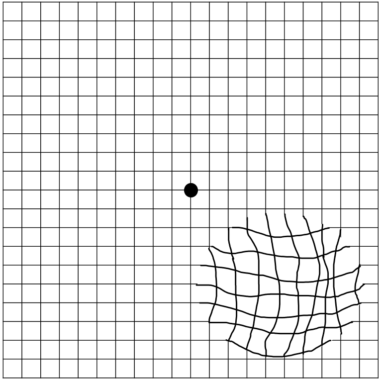
Patient will present as → a 62-year-old male who arrives for his follow-up visit for chronic, gradual central vision loss. He describes a phenomenon of wavy or distorted vision that has deteriorated rather quickly. The patient is frustrated because he "just can't drive anymore,” and he is "having difficulty seeing words when he reads.” When looking at a specific region of the Amsler grid, he reports a dark “spot” in the center, with bent lines. On the fundoscopic exam, you note areas of retinal depigmentation along with the presence of yellow retinal deposits.


To watch this and all of Joe Gilboy PA-C's video lessons you must be a member. Members can log in here or join now.
Gradual PAINLESS loss of CENTRAL vision (versus glaucoma, which presents with a peripheral → central loss)
- The macula is responsible for central visual acuity, which is why macular degeneration causes gradual central field loss
- Macular Degeneration is the most common cause of permanent legal blindness and visual loss in the elderly > 75
- The most important risk factors for age-related macular degeneration are increasing age, cigarette smoking, and family history (genetic predisposition), with hypertension and hyperlipidemia as major secondary contributors.
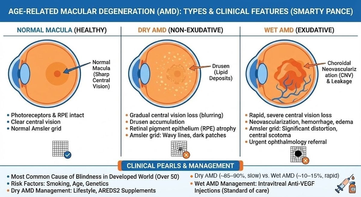
- Dry macular degeneration (85% of cases): atrophic changes with age – slow, gradual breakdown of the macula (macular atrophy), with DRUSEN (DRY) = yellow retinal deposits
- Wet macular degeneration: an advanced form of dry age-related macular degeneration. New blood vessels growing beneath the retina (neovascularization) leak blood and fluid, damaging the retinal cells. These small hemorrhages usually result in rapid and severe vision loss

"Dry AMD progresses to wet AMD when drusen-induced retinal hypoxia triggers VEGF-mediated choroidal neovascularization, leading to subretinal bleeding and rapid central vision loss → Wet AMD is what happens when dry AMD becomes ischemic and inflammatory enough to trigger abnormal blood vessel growth!"
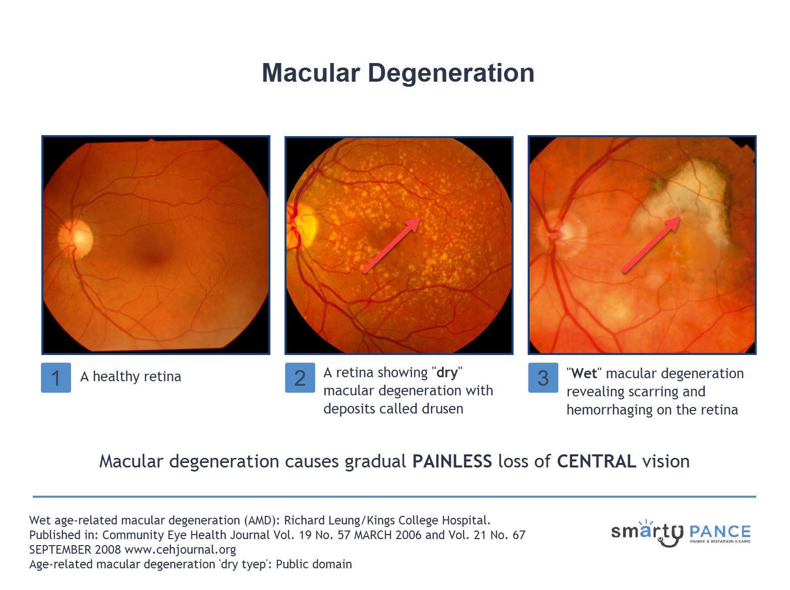
Dilated funduscopic findings are diagnostic; color photographs, fluorescein angiography, and optical coherence tomography assist in confirming the diagnosis and in directing treatment.
- There is no USPSTF-recommended population screening for AMD, but routine dilated ophthalmologic exams and Amsler grid monitoring are recommended in at-risk and diagnosed patients.
- The American Academy of Ophthalmology (AAO) recommends routine dilated eye exams:
- Age ≥40 → baseline dilated exam
- Age ≥65 → every 1–2 years
- The American Academy of Ophthalmology (AAO) recommends routine dilated eye exams:
Wet - Age-Related Macular Degeneration - hemorrhage or fluid in the subretina
- Neovascularization: macular grayish-green discoloration

Dry - Age-Related Macular Degeneration
- Drusen deposition
- areas of retinal atrophy (depigmentation)
- retinal pigment epithelium mottling (pigmentation)
Amsler grid: line distortion seen on the grid
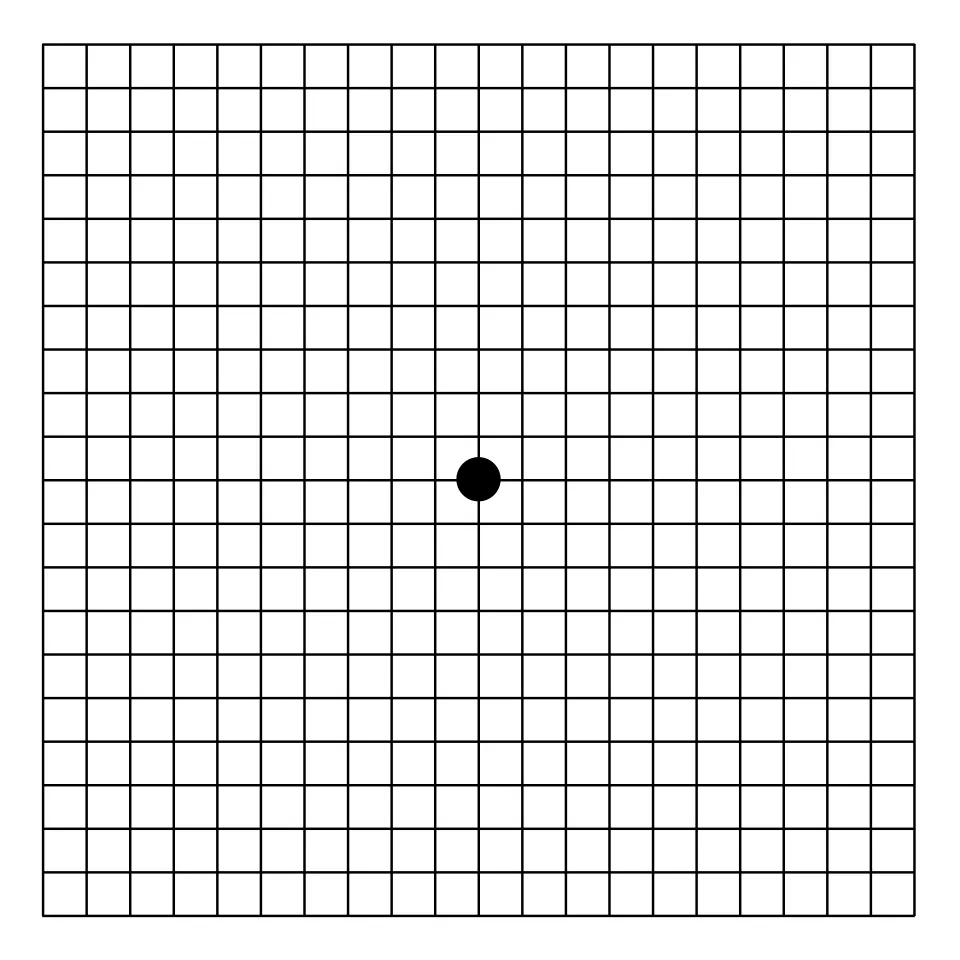
Normal Amsler Grid. Image: Jmarchn, CC BY-SA 3.0, via Wikimedia Commons.
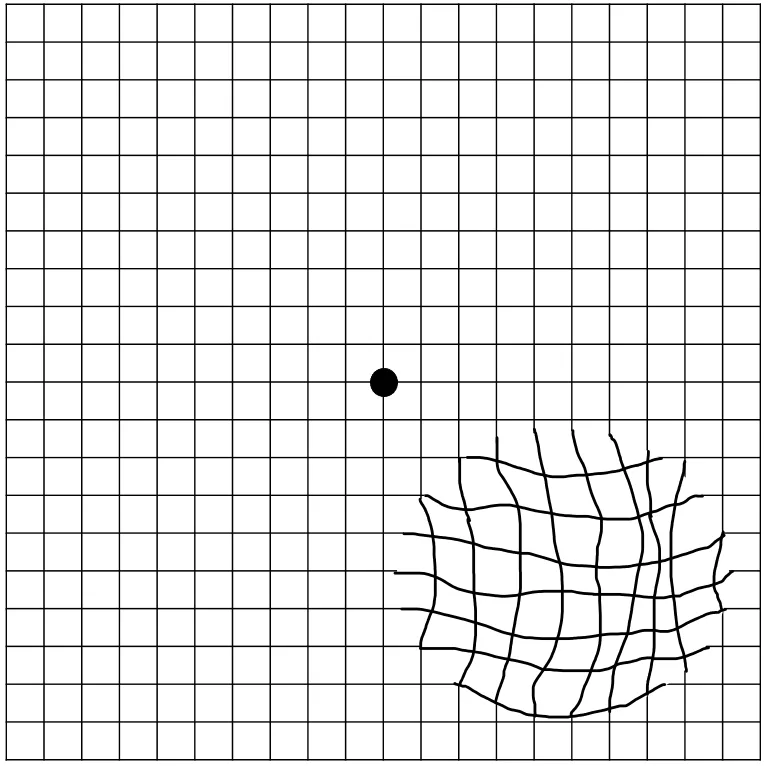
Amsler grid as it might be viewed by a person with age-related macular degeneration. Image: Isislunsky, CC BY-SA 4.0, via Wikimedia Commons.
Wet Age-Related Macular Degeneration
- VEGF inhibitors (e.g., bevacizumab)
- Photodynamic therapy
- Zinc and antioxidant vitamins
Dry Age-Related Macular Degeneration
- Zinc and antioxidant vitamins

Age-related macular degeneration (AMD) is the most common cause of irreversible central vision loss. It affects patients over 60 years of age and is related to retinal aging. There are two types: dry (nonexudative) and wet (exudative). Dry AMD accounts for the majority of the diagnoses of the condition, while wet AMD is the more severe form. Prolonged exposure to UV light, smoking, lighter colored eyes, and hyperopia are risk factors.
Play Video + QuizQuestion 1 |
Night vision Hint: Night vision is a function of rod photoreceptors, which are found in the peripheral retina. | |
Color vision Hint: Color vision is a function of cone photoreceptors | |
Peripheral vision Hint: The peripheral retina is responsible for peripheral vision. | |
Central vision acuity |
Question 2 |
Dry (atrophic) age-related macular degeneration | |
Primary open-angle glaucoma Hint: Glaucoma takes peripheral vision first and causes optic disc cupping, not drusen. | |
Nuclear sclerotic cataract Hint: A cataract clouds the lens. It doesn't put yellow deposits under the retina. | |
Central retinal vein occlusion Hint: Central retinal vein occlusion causes sudden vision loss with widespread retinal hemorrhages. | |
Retinal detachment Hint: Retinal detachment causes sudden, curtain-like vision loss, often with floaters and flashes. |
Question 3 |
Blepharitis Hint: Blepharitis is a chronic bilateral inflammatory condition of the lid margins | |
Cataracts Hint: Cataracts are the clouding of the lens sufficient to reduce vision. Most develop slowly as a result of aging, leading to gradual impairment of vision. | |
Central retinal artery occlusion Hint: Central retinal artery occlusion presents as a rare cause of sudden profound monocular visual loss. | |
Macular degeneration |
Question 4 |
High-dose oral beta-carotene (vitamin A) Hint: Supplements with lutein and zeaxanthin (not beta-carotene) only slow progression. They don't treat active wet AMD. | |
Intravitreal anti-VEGF injection | |
Topical timolol Hint: Timolol lowers eye pressure for glaucoma. | |
Oral prednisone Hint: Systemic steroids don't make the new vessels regress. | |
Observation, since macular degeneration can't be treated Hint: Wet AMD is treatable, and waiting lets central vision get worse. |
|
List |
References: Merck Manual · UpToDate