The NCCPA™ PANCE EENT Content Blueprint => disorders of the eye => vascular disorders
| Central Retinal Artery Occlusion (CRAO) | Central Retinal Vein Occlusion (CRVO) | |
| Pathophysiology | Supply Problem: Embolism or thrombosis leads to retinal ischemia (an "eye stroke"). | Drainage Problem: Thrombus in the central retinal vein causes back pressure and congestion. |
| Presentation | Sudden, profound, painless monocular vision loss. Often permanent. | Sudden or subacute, painless monocular vision loss. Variable severity. |
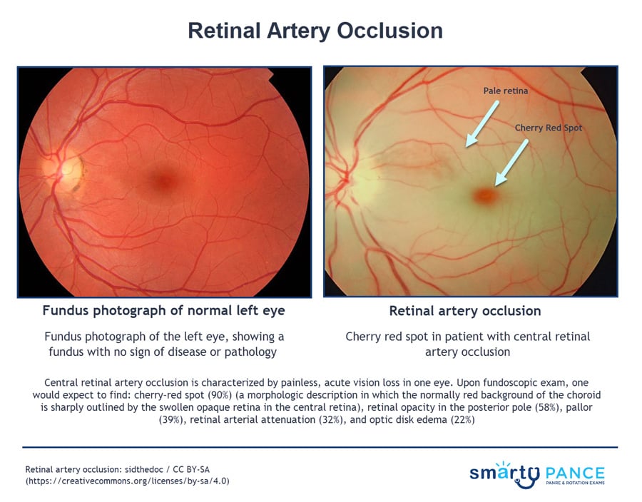
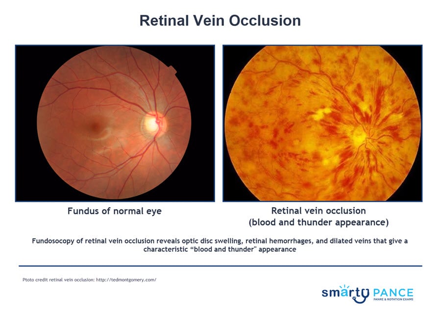
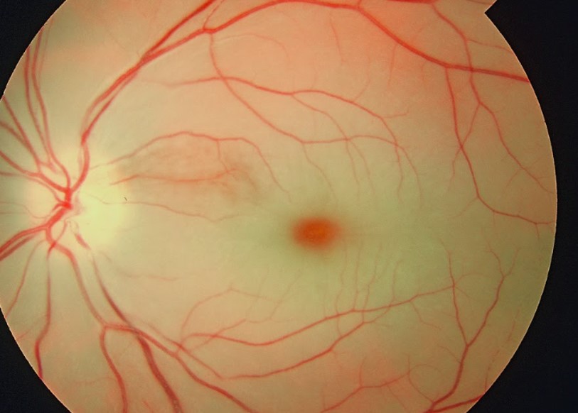
| Fundoscopic Findings | Pale, edematous retina with a cherry-red spot (macula) and "box-carring" of vessels. | "Blood and thunder" appearance: diffuse hemorrhages, optic disc edema, and tortuous veins. |
| Primary Risk Factors | Carotid artery disease, A-fib, temporal arteritis (GCA), and atherosclerosis. | Hypertension, diabetes, glaucoma, and hypercoagulable states. |
| Management Goals | Emergent: Decrease IOP (acetazolamide, ocular massage) to dislodge the clot; rule out GCA. | Consultation: Manage macular edema (anti-VEGF) and prevent neovascular glaucoma. |
| PANCE Trigger | Pale retina + Cherry-red spot. | Blood and thunder + Disc edema. |
| Retinal vascular occlusion | Patient will present as → a 74-year-old man with sudden vision loss in his right eye. He has a medical history of hypertension, coronary artery disease, and new-onset atrial fibrillation. On physical exam, a carotid bruit is auscultated. His visual acuity is light perception. Confrontational visual fields reveal a dense scotoma, and a penlight examination shows an afferent pupillary defect. Dilated funduscopic examination shows retinal whitening with a cherry-red spot in the fovea. Central retinal artery occlusion (cherry-red spot, ischemic retina)
DX: Fundoscopy
TX: Emergent ophthalmologic consult - Immediate treatment is indicated if occlusion occurs within 24 hours of presentation
Patient will present as → a 65-year-old man with a history of hypertension and hyperlipidemia presents to the emergency department complaining of unilateral sudden, painless vision loss in his right eye that started 2 hours ago. On examination, his visual acuity in the right eye is 20/200, and fundoscopy reveals retinal hemorrhages, dilated and tortuous retinal veins, and cotton-wool spots. There is no evidence of neovascularization. Central retinal vein occlusion (blood and thunder fundus)
DX: Funduscopy: retinal hemorrhages in all quadrants, optic disc swelling; blood and thunder retina (dilated veins, hemorrhages, edema, exudates) TX: vision resolves with time (partially); workup for thrombosis
|