Patient with dacryoadenitis will present as → a 32-year-old woman presents to the emergency department with a 2-day history of pain, redness, and swelling in the outer corner of her right eye. She denies any vision changes, discharge, or trauma. She has no significant past medical history and is not on any medications. On physical examination, you note localized erythema and swelling over the lateral aspect of her right upper eyelid. Her visual acuity is normal, and there is no proptosis.
Patient with dacryocystitis will present as → a 58-year-old woman presents to the emergency department with a 3-day history of increasing pain, redness, and swelling in the inner corner of her left eye. She also reports some purulent discharge from the same eye. She denies any vision changes or trauma. On physical examination, you note localized erythema, warmth, and swelling over the medial canthal area of her left eye. Her visual acuity is normal.
Lacrimal gland vs. lacrimal sac vs. lacrimal duct
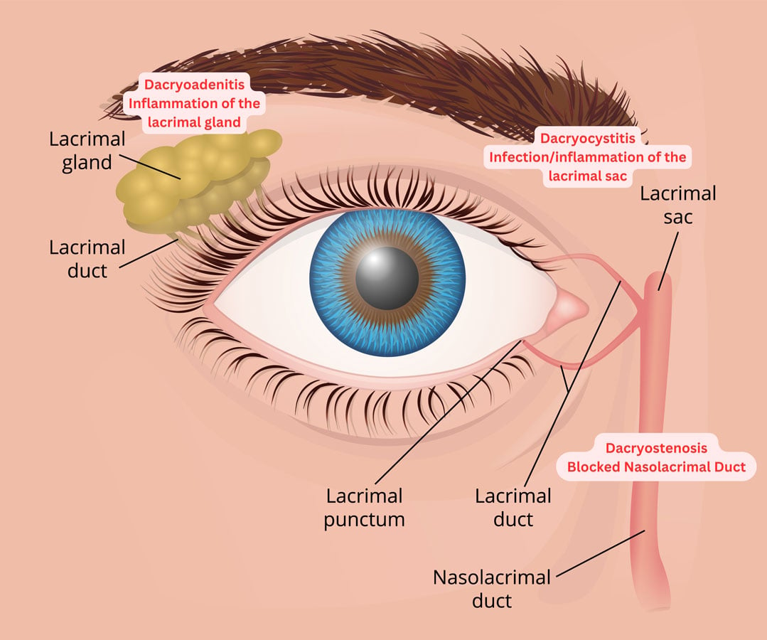
Image © by Adobe Stock (with edits by Smarty PANCE)
The lacrimal gland makes tears, while the lacrimal sac temporarily stores tears (made in the lacrimal gland), preventing them from constantly flooding the lacrimal ducts. The lacrimal duct, or the nasolacrimal duct, drains tears into the nasal cavity.
Dacryoadenitis is an infection or inflammation of the lacrimal (tear-producing) gland, commonly caused by infection (bacterial or viral) or systemic inflammatory conditions (supratemporal)
- s/sx: unilateral severe pain, swelling, redness, tearing, drainage
- Acute dacryoadenitis is most commonly due to viral or bacterial infection. Common causes include mumps, Epstein-Barr virus, staphylococcus, and gonococcus.
- Chronic dacryoadenitis is most often due to noninfectious inflammatory disorders. Examples include sarcoidosis, thyroid eye disease, and orbital pseudotumor
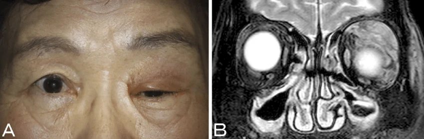 Dacryoadenitis presenting as an erythematous, swollen upper eyelid and ptosis. From "Dacryoadenitis with Ptosis and Diplopia as the Initial Presentation of Granulomatosis with Polyangiitis" by Makoto Hibino and Tetsuri Kondo via ResearchGate. Licensed under CC BY-NC-ND 4.0.

"On the boards you may be able to differentiate between the two based on the location: dacryocystitis will be medial. Remember, medial = Center = dacroCystitis. vs. dacryoAdenitis, which is up, up, and Away."
Dacryocystitis is an infection or inflammation of the lacrimal sac, typically caused by obstruction of the nasolacrimal duct (inferomedial region)
- The lacrimal sac's function is to temporarily store tears that drain from the eyes and prevent them from constantly flooding the tear ducts
- Dacryocystitis will be medial (remember "c" for central)
- Common causes include Staphylococcus aureus, Streptococcus species, and, less commonly, Gram-negative bacteria
- Risk factors include nasolacrimal duct obstruction, sinus infections, and trauma to the lacrimal system
- Symptoms include pain, swelling, erythema, and tenderness over the inner aspect of the lower eyelid near the lacrimal sac, often accompanied by tearing (epiphora) and discharge

Dacryocystitis with swelling and inflammation over the lacrimal sac. Photo by Natanalyzator via Wikimedia Commons, CC BY-SA 4.0.
The diagnosis of dacryocystitis and dacryoadenitis based on clinical observation
- In cases of chronic dacryoadenitis/dacryocystitis, a CT or MRI of the orbits may be warranted
- In acute cases of dacryocystitis, a tear duct massage can be performed to express material for culture and gram stain
- In patients who appear to be acutely toxic or those who present with visual changes, imaging and bloodwork should be considered
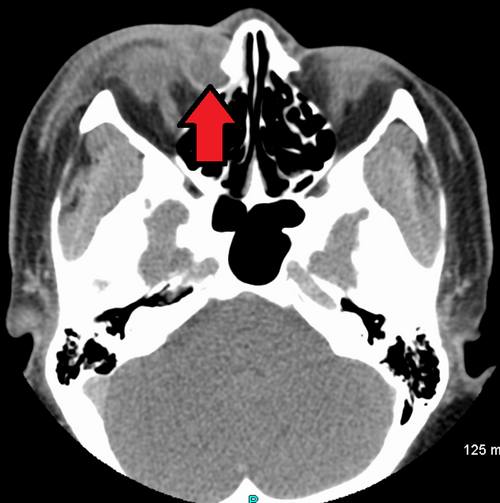
CT showing dacryocystitis with inflammation of the lacrimal sac adjacent to the medial orbit (arrow). Photo by James Heilman, MD via Wikimedia Commons, CC BY-SA 4.0.
Dacryoadenitis
Acute treatment involves:
-
- Antibiotics for bacterial causes (e.g., cephalexin or amoxicillin-clavulanate)
- Supportive care for viral causes (e.g., warm compresses, analgesics)
- Chronic treatment focuses on managing the underlying systemic condition
- Surgical drainage may be needed for abscess formation, and biopsy may be required in chronic or atypical cases to exclude malignancy
- Complications include lacrimal gland abscess, vision impairment, or progression to orbital cellulitis
Dacryocystitis
- Warm compresses and gentle massage may help alleviate symptoms
- Acute dacryocystitis (< 3 months) requires oral antibiotics (e.g., cephalexin or amoxicillin-clavulanate) for mild cases and IV antibiotics for severe infections or systemic involvement
- Chronic dacryocystitis (> 3 months) typically presents with fewer inflammatory signs and requires surgical therapy for the underlying cause
- Definitive treatment involves addressing the underlying obstruction, often with dacryocystorhinostomy (DCR), a surgical procedure to restore tear drainage
- Complications include abscess formation, orbital cellulitis, and, rarely, sepsis
Question 1 |
A 7-month-old female is brought to your clinic by her mother, who is concerned about persistent swelling at the nasal corner of the child's left eye. The mother reports that the swelling has been present for several weeks and occasionally becomes red and inflamed. If this condition is left untreated, which of the following complications is most likely to develop?
Hyphema Hint: This refers to the presence of blood in the anterior chamber of the eye, typically resulting from trauma or eye surgery, not related to nasolacrimal duct obstruction. | |
Papilledema Hint: This is swelling of the optic disc due to increased intracranial pressure and is not a complication associated with nasolacrimal duct obstruction. | |
Pterygium Hint: A pterygium is a benign growth on the conjunctiva that can encroach onto the cornea, usually related to UV light exposure, not nasolacrimal duct issues. | |
Dacryocystitis | |
Orbital Cellulitis Hint: While severe untreated eye infections can potentially lead to orbital cellulitis, it is less likely in this scenario without evidence of a more significant or spreading infection. |
Question 2 |
Location of swelling: Dacryoadenitis - lateral canthus, Dacryocystitis - medial canthus. | |
Discharge: Dacryoadenitis - mucopurulent, Dacryocystitis - purulent. Hint: While both can have purulent discharge, it's not a reliable differentiator. | |
Tenderness: Dacryoadenitis - absent, Dacryocystitis - present upon lacrimal sac palpation. Hint: Both conditions can have tenderness, though lacrimal sac palpation in Dacryocystitis might be more pronounced. | |
Fever: Dacryoadenitis - common, Dacryocystitis - rare. Hint: Fever can be present in both, especially with infections. | |
Lacrimation: Dacryoadenitis - decreased, Dacryocystitis - increased. Hint: Dacryoadenitis typically presents with decreased tear production, while Dacryocystitis can have either increased or decreased tears depending on the cause and obstruction. |
Question 3 |
Intravenous antibiotics | |
Warm compresses and continued oral antibiotics Hint: Appropriate for mild cases but not for worsening symptoms. | |
Dacryocystorhinostomy Hint: A surgical procedure indicated for chronic dacryocystitis or obstruction, not acute infection. | |
Topical corticosteroids Hint: Not indicated in acute infections and can potentially worsen the condition. | |
Incision and drainage of the lacrimal sac Hint: Typically reserved for abscess formation or severe cases not responding to intravenous antibiotics. |
Question 4 |
Intravenous antibiotics | |
Warm compresses and continued oral antibiotics Hint: Appropriate for mild cases but not for worsening symptoms. | |
Dacryocystorhinostomy Hint: A surgical procedure indicated for chronic dacryocystitis or obstruction, not acute infection. | |
Topical corticosteroids Hint: Not indicated in acute infections and can potentially worsen the condition. | |
Incision and drainage of the lacrimal sac Hint: Typically reserved for abscess formation or severe cases not responding to intravenous antibiotics. |
|
List |
References: Merck Manual · UpToDate