NCCPA™ PANCE EENT Content Blueprint ⇒ ear disorders ⇒ middle ear
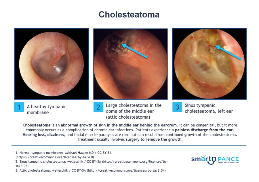
| Cholesteatoma | Patient will present as → a 43-year-old male with a “lifelong” history of chronic ear infections and episodic purulent drainage from his right ear canal. The patient is currently without symptoms. Examination of the ear shows a clear external canal, but the tympanic membrane is retracted, and there is a pocket of white material and an opacity of the pars flaccida. The Weber test lateralizes to the right, and Rinne shows air conduction > bone conduction on the left and bone conduction > air conduction on the right. Preparations are made to undergo a non-contrast computed tomography (CT) scan of the temporal bone. Cholesteatoma is an abnormal growth of squamous epithelium in the middle ear or mastoid that can lead to destruction of nearby structures
DX: Diagnosis can be made with otoscopic visualization revealing a retraction pocket or a mass of keratin debris
TX: Surgical removal to prevent complications and restore hearing
|
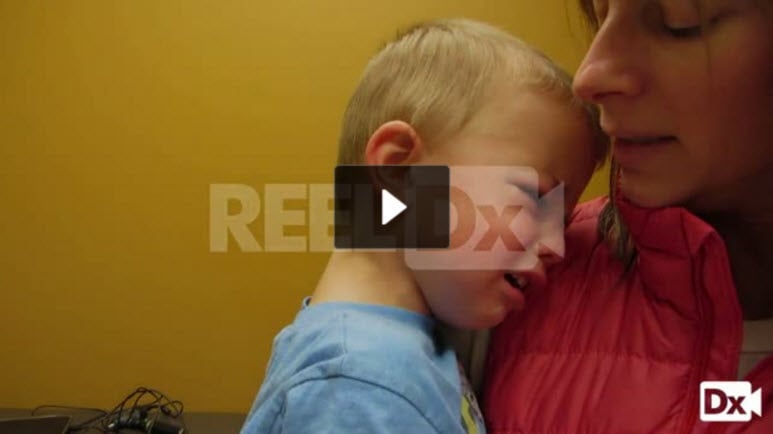
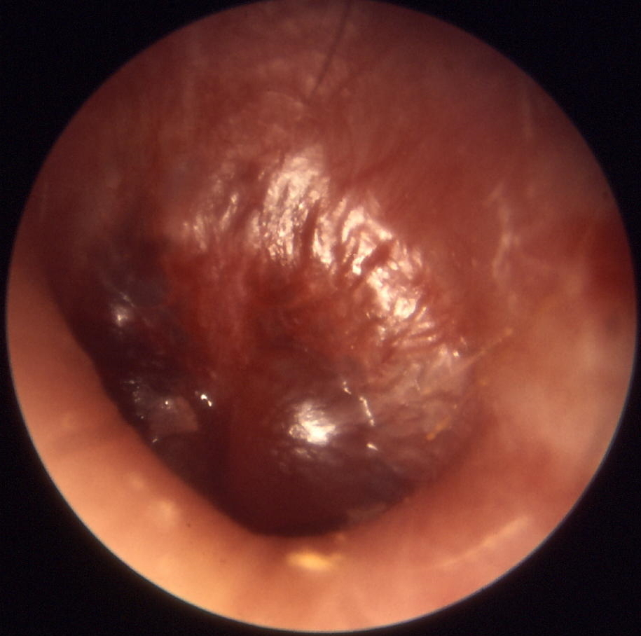
| Otitis media (ReelDx) | ReelDx Virtual Rounds (Otitis media)Patient will present as → a 3-year-old previously healthy male is brought to your office by his mother. The mother reports the child has been crying and pulling at his right ear over the past 2 days and reports the patient has been febrile the past 24 hours. The patient’s past medical history is unremarkable, although the mother reports the patient had a “common cold” a week ago, which resolved without intervention. His temperature is 101.6 F, blood pressure is 100/70 mmHg, pulse is 120/min, and respirations are 22/min. The otoscopic exam is seen here. Acute otitis media (AOM) is a bacterial infection of middle ear fluid that typically follows a viral URI, peaking at 6–24 months. Diagnosis requires middle ear effusion + acute inflammation: a bulging TM, new otorrhea, or mild bulging with acute otalgia or intense erythema.
DX: Otoscopy — bulging, loss of landmarks, erythema, injection of the TM, and purulent effusion
TX: High-dose amoxicillin 90 mg/kg/day divided BID (max 4 g/day) — 10 days if < 2 years, 5–7 days if ≥ 2 years; observation for 48–72 hours is acceptable for nonsevere unilateral disease in children ≥ 6 months
 Acute otitis media - Note bulging, loss of landmarks, redness, injection of TM, and pus. Photo by Michael Hawke, MD via Wikimedia Commons, CC BY 4.0. |
| Otosclerosis | Patient will present as → a 35-year-old woman presents with a 2-year history of progressive hearing loss in both ears. She notes that her hearing is worse in noisy environments and denies any history of ear infections, trauma, or exposure to ototoxic medications. Her mother also experienced similar hearing loss in her 30s. On physical examination, the tympanic membranes appear normal bilaterally. An audiogram reveals conductive hearing loss with a Carhart notch at 2000 Hz. A CT scan is performed, demonstrating bony changes around the otic capsule, particularly near the stapes footplate. Otosclerosis is a progressive disorder characterized by abnormal bone remodeling in the middle ear, leading to conductive hearing loss.
DX: Diagnosed with audiometry, showing conductive hearing loss with a normal tympanogram. CT scan of the temporal bone may show bony changes TX: Treatment options include hearing aids for amplification and surgical stapedectomy or stapedotomy to improve hearing 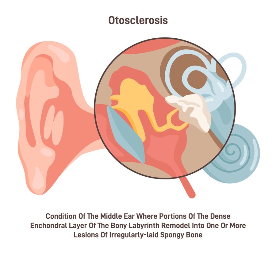 © inspiring.team / Adobe Stock |
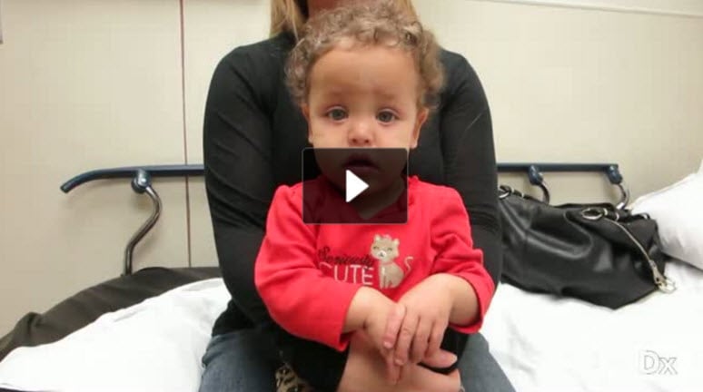
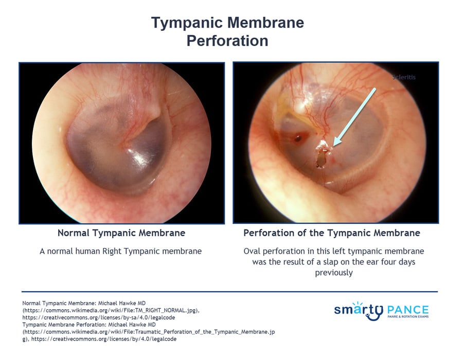
| Tympanic membrane perforation (ReelDx) | ReelDx Virtual Rounds (Tympanic membrane perforation)Patient will present as → a 9-month-old female with nasal congestion and cough is brought to your clinic by her mother, who reports that the child is very fussy, has been tugging at her right ear, and refuses to eat. On physical exam, you note copious green/yellow nasal discharge and right-sided otorrhea. An otoscopic exam reveals a significant amount of clear/white discharge obstructing your view. With careful examination, you are able to observe a ruptured right tympanic membrane. Tympanic membrane perforation is a tear in the eardrum caused most often by acute otitis media (pressure necrosis) or trauma (cotton swab, slap to the ear, barotrauma, blast). It presents with otalgia, otorrhea, and conductive hearing loss.
DX: Otoscopy visualizes the defect — conductive hearing loss on tuning forks (Weber lateralizes to the affected ear, Rinne bone > air)
TX: Most heal spontaneously within weeks (a perforation < 25% of the drum usually closes by 4 weeks) — keep the ear dry (no swimming; petroleum-jelly cotton ball when bathing)
|