


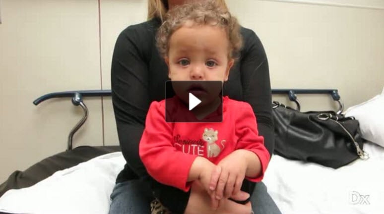
9 m/o with fussiness and decreased oral intake
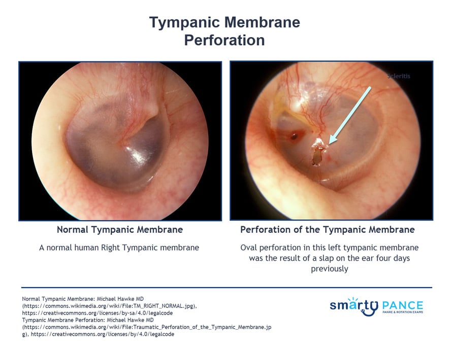
Patient will present as → a 9-month-old female with nasal congestion and cough is brought to your clinic by her mother, who reports that the child is very fussy, has been tugging at her right ear, and refuses to eat. On physical exam, you note copious green/yellow nasal discharge and right-sided otorrhea. An otoscopic exam reveals a significant amount of clear/white discharge obstructing your view. With careful examination, you are able to observe a ruptured right tympanic membrane.
- AOM perforation — purulent otorrhea with abrupt relief of pain as middle ear pressure decompresses
- Traumatic perforation — sudden severe pain, sometimes bloody otorrhea, hearing loss, tinnitus; ⚠️ vertigo, nystagmus, vomiting, or facial weakness = inner ear or ossicular injury
- Most heal spontaneously — a perforation < 25% of the drum usually closes within 4 weeks; most are uncomplicated with hearing loss < 40 dB and no vestibular complaints
- Location matters — central perforations are benign; marginal/attic perforations or margins folded into the middle ear carry cholesteatoma risk
- Water precautions until healed — no swimming; petroleum-jelly cotton ball when showering; swimmers and divers should have persistent perforations repaired for a "safe ear"
Otoscopy visualizes the defect — do not irrigate the canal, and in trauma with other signs of middle ear injury, leave canal blood clots and foreign bodies for ENT
- Defer pneumatic otoscopy when the TM is perforated — it can push air into an injured otic capsule and provoke vertigo
- Tuning forks — Weber lateralizes to the injured ear = conductive loss (away = sensorineural); Rinne bone > air ≈ conductive loss > 40 dB → suspect ossicular disruption if > 50–60 dB
- Audiometry within 24 hours for every traumatic perforation — hearing loss ≥ 40 dB signals serious middle ear injury
- Fine-cut temporal bone CT (not a routine head CT) if basilar skull fracture signs (hemotympanum, Battle sign, CSF otorrhea), facial nerve palsy, hearing loss ≥ 40 dB, or vestibular symptoms
- Water precautions and pain control; no antibiotics for a clean perforation
- Ofloxacin otic drops (5 drops BID × 3–5 days) only if the wound is contaminated (water, external puncture) or the canal is occluded with blood or drainage
- If drops are used, choose a fluoroquinolone (ofloxacin or ciprofloxacin-dexamethasone) — non-ototoxic; never aminoglycoside drops (neomycin/Cortisporin) through a perforated TM
- Audiometry within 24 hours, then primary care + audiology follow-up at 4 weeks to confirm closure and hearing recovery
- Not healed or hearing not recovered at 4 weeks → ENT; persistent perforations (typically > 3 months) need tympanoplasty
AOM with perforation — oral amoxicillin 90 mg/kg/day × 10 days; topical drops alone are not appropriate for a spontaneous AOM perforation
⚠️ Emergency ENT evaluation within 48 hours for hearing loss ≥ 40 dB (or decreased perception of normal speech), vestibular signs (nystagmus, vomiting, ataxia), basilar skull fracture, CSF otorrhea, or facial nerve injury
- Ossicular disruption → surgical repair within 48 hours for best hearing outcome
- Immediate facial paralysis → urgent surgical exploration; delayed paralysis (> 1 day) is nerve edema → IV glucocorticoids
- Contaminated middle ear wounds are tetanus-prone; unexplained ear trauma in a young child or older adult → consider abuse
Question 1 |
Tobramycin otic drops Hint: While effective against Pseudomonas, tobramycin can be ototoxic and is generally avoided if there is a possibility of tympanic membrane perforation. | |
Erythromycin solution Hint: Erythromycin is not typically the first choice for otitis externa, as it has a narrower spectrum of activity against the common pathogens. | |
Offloxacin otic drops | |
Gentamicin drops Hint: Gentamicin is also effective against Pseudomonas but carries a risk of ototoxicity and is not preferred if tympanic membrane status is uncertain. | |
Acetic acid otic solution Hint: Acetic acid solution can be used for mild cases of otitis externa but may not be sufficient for more severe infections, especially when there is significant swelling and discharge. |
Question 2 |
Acute otitis media | |
Foreign body insertion Hint: While foreign bodies can cause TM perforation, especially in children, they are less common than perforations due to AOM. | |
Barotrauma Hint: Barotrauma, such as from diving or flying, can lead to TM perforation, but it is less common compared to AOM. | |
Chronic otitis media Hint: Chronic otitis media can lead to TM perforation, but acute otitis media is a more frequent cause. | |
Loud noise exposure Hint: Exposure to loud noises, such as explosions or gunfire, can cause TM perforation, but these instances are less common compared to AOM. |
Question 3 |
Immediate referral for audiometry Hint: Useful for assessing the extent of hearing loss but not urgent. | |
Prescription of oral antibiotics Hint: Not routinely indicated unless there is evidence of infection. | |
Keep the ear dry and reevaluate in a few weeks | |
CT scan of the temporal bone Hint: Indicated if there is suspicion of more extensive injury. | |
Myringotomy Hint: A surgical procedure not typically required for traumatic tympanic membrane perforation. |
Question 4 |
Continued observation Hint: Not appropriate given the duration and persistent symptoms. | |
Tympanoplasty | |
Systemic corticosteroids Hint: Not effective for tympanic membrane perforation. | |
Topical antibiotic drops Hint: Used to prevent infection but do not facilitate healing of the perforation. | |
Hearing aid fitting Hint: May be considered for permanent hearing loss but after addressing the perforation. |
|
List |
References: Merck Manual · UpToDate